WHAT’S IN A CELL? #3
Hello, readers. In order to study the function of a particular
organelle it is often helpful to isolate it from the rest of the cell. This can
be done by cell fractionation, as briefly outlined below:
- A Liver tissue is placed in a fluid that is an ice-cold isotonic buffer. When the cell is broken up, membranes are ruptured and many compounds that do not normally mix are brought together. The low temperature minimizes unwanted reactions, including self-digestion by digestive enzymes. The isotonic solution prevents organelles gaining or losing water by osmosis, and the buffer prevents any changes in pH.
- Tissue is cut into small pieces.
- Tissue is put into blender/homogeniser to break up whole cells.
- Mixture filtered to remove debris.
- Filtrate spun in centrifuge.source
Principle: The organelles will separate out in a particular order, according to their density and shape. After a time, the sediment containing a particular type of organelle can be separated from the supernatant (the liquid that contains the remaining organelles). The exact times vary from tissue to tissue.source
Size and scaling – getting a sense of proportion
An essential part of microscopy is to know the actual size of the objects being studied. There are only three units of information to work with.
- The actual size of the object.
- The magnification.
- The size of the image on the paper or screen
If you know two of these values, you can work out the third one. The formula is:
Magnification = Observed size ÷ Actual size
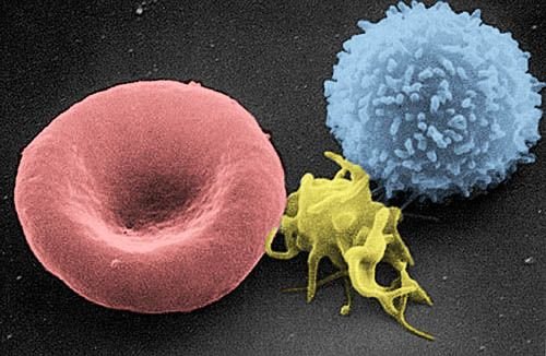
Often an exam question will show a micrograph or diagram, give you the magnification and ask you to work out the actual size.Take, for instance, in the coloured scanning electron micrograph of red blood cells, white blood cells and bacteria (a type of staphylococcus) that’s having a magnification of ×7500. The image is ×7500 times larger than the cell really is.
Measuring the diameter of the bottom red blood cell, we find it is 40mm. Converting mm into micrometres, 40 mm is 40000μm. The real cell must be 7500 times smaller than that. So actual size is 40000/7500 = 5.3 μm.source
Recognize a silly answer when you see one
It’s a great help if you know the sort of answer you are looking for. If you measure a bacterium and calculate it to be 3 mm long, there’s something wrong somewhere.
Remember the golden rules:
1 mm = 1000 μm (micrometres)
1 μm = 1000 nm (nanometres)
The following are useful guidelines
- Animal cells are usually 20-50 μm in diameter, while plants cells are usually larger, perhaps 100-150 μm.
- Organelles such as nuclei and mitochondria are obviously smaller. An average nucleus may be 10 μm in diameter while mitochondria range from 1-10 μm.
- Most bacteria are much smaller than plant and animal cells. An average bacterium would be about the same size as a mitochondrion (there may be a good reason for this which I’ll explain further in the endosymbiont theory). The largest known bacteria are 250 μm long but the smallest are just 0.3 μm long.
- Small organelles and large molecules are measured in nm. The plasma membrane, for example, is only about 8 nm thick.
- Viruses are really, really tiny. The largest ones measure about 400 nm – slightly larger than the smallest known bacteria. The smallest viruses are just 20 nm in diameter.source
Autoradiography
Electron microscopes are useful tools for investigating cell structure, but they don’t reveal much about the processes going on inside a living cell. After World War II, work on the atomic bomb produced chemicals called radioactive isotopes as a by-product. These are very useful for studying metabolism because they behave in the same way as non-radioactive atoms and their movements through the cell can be traced.
After cells are exposed to a radioactive isotope such as carbon-14, they are washed to remove excess isotopes and then fixed, embedded, sectioned and mounted on microscope slides. The slides are then taken to a darkroom and coated with photographic emulsion that has been heated to make it melt. After coating, the slides are cooled and kept in the dark for various time periods from a few days to several weeks. During this time, radioactive decay causes a chemical change to occur in the film of photographic emulsion. When this is developed using standard photographic techniques and the slide is examined under the light microscope, silver grains can be seen in the areas exposed by the radioactivity. The pattern of grains shows the structures of the cells that have incorporated the radioactive label.source
EUKARYOTES: PLANT CELLS
In a typical leaf cells from a plant, as seen with the light microscope, there are palisade mesophyll cells. Plant cells have several features that are not found in animal cells. A plant cell is surrounded by a cell wall made of cellulose. A large proportion of the inside of the cell is taken up with a fluid-filled compartment known as the vacuole. Together, the cell wall and vacuole maintain the shape of the whole cell.
Plant cells have specialized organelles, the chloroplasts, which enable them to make their own food by photosynthesis. Chloroplasts bring together all the chemicals involved in photosynthesis, and provide sites for the chlorophyll molecules so that they can absorb the maximum amount of light.source
THE CELLULOSE CELL WALL
Unlike animal cells, plant cells are enclosed by a cell wall that consists of many cellulose fibres cemented together by a mixture of other organic substances.
Cellulose is a polysaccharide (a sugar polymer) which consists of long straight chains of glucose molecules bonded to adjacent molecules by hydrogen bonds. In the cell wall, around 2000 parallel cellulose molecules are packed together to form microfibrils. These in turn are bundled together to form macrofibrils. Like fibreglass, the cell wall has great strength because of the many strong fibres and the ‘glue’ that holds them together.
The main functions of the cell wall are:
- to provide rigidity and strength to the cell. Cell walls need to be strong enough to resist expansion to allow the cell to become turgid;
- to force the cell to grow in a certain way. A particular arrangement of fibrils causes the cell to assume a particular shape – for example, a long, thin tube.source
VACUOLES
Plant cells have a large, fluid-filled cavity called a vacuole. In mature cells the vacuole can occupy over 80 per cent of the cell volume, and is filled with a fluid called cell sap. The sap consists of a complex mixture of sugars, salts, pigments and waste products in water.
Vacuoles have several functions:
- They absorb water by osmosis and therefore swell, pushing the cytoplasm against the cell wall. In this state, a plant cell is said to be turgid.
- They store food substances such as sugars and mineral salts.
- They store pigments that give colour to plant structures such as petals.
- They can accumulate waste products and by-products of metabolism. Sometimes these chemicals may be toxic or have an unpleasant taste and are used by the plant to make it less palatable to herbivores.source
CHLOROPLASTS AND OTHER PLASTIDS
Chloroplasts Are one of a group of plant cell organelles known as plastids. Chloroplasts are surrounded by a double membrane and contain an elaborate internal membrane system that houses the chemicals of photosynthesis.source
THE ORIGIN OF MITOCHONDRIA AND CHLOROPLASTS: The Endosymbiont Theory.
Scientists continue to speculate whether or not mitochondria and chloroplasts originated from outside eukaryotic cells. Many researchers think that they were originally free-living prokaryotes, similar to bacteria and blue-green bacteria, which, at some point, became incorporated into larger cells.source
Let’s look at the evidence, mitochondria and chloroplasts have several similarities to prokaryotes:
- They are similar in size to prokaryotes.
- They have their own DNA, as prokaryotes do, suggesting an ability to reproduce independently.
- The DNA of both organelles is circular, like that of bacteria.
- Both organelles have their own ribosomes, smaller than those in eukaryotic cells but identical to those in prokaryotes.
- All reproduce by binary fission.
- The biochemistry of mitochondria is closer to that of aerobic bacteria than that of the eukaryotic cell; chloroplasts and blue-green bacteria are even more similar.source
This theory was first suggested by team led by Lyn Margulis at Boston University, USA. Assuming he theory is true, how could these organisms have become incorporated into a larger cell without being destroyed by lysosomes?source
Imagine a heterotrophic, anaerobic organism – similar to an amoeba in structure and movement – that feeds by phagocytosis and respires without oxygen, though oxygen is present in its environment. If such a cell were to ingest aerobic bacteria that it did not digest, the bacteria could respire aerobically, and in doing so could provide the host organism with far more ATP than the host could produce on its own. The host cell in turn would give the bacteria material to respire.
Similarly chloroplasts, which may originally have been free-living blue green bacteria, could also have become incorporated into larger cells. If these were ingested but not digested – they would continue to photosynthesize within the host and so provide it with organic food and oxygen – an obvious advantage. The host, in turn, could provide the blue-green bacterium with carbon dioxide and nitrogen.
Associations in which an autotroph lives inside a heterotroph, or an aerobic lives inside an anaerobic, are both very convenient arrangements. An autotroph is an organism that can make its on food: a green plant is an autotroph that makes its own food by photosynthesis. A heterotroph is an organism that must obtain ready- made food: a cow is a heterotroph that obtains its food from grass.
One organism produces what the other needs. This is the key to the success of several other associations, for example, lichens (algae and fungi) and corals (algae inside animals). Both host and ‘passenger’ gain a survival advantage: the host gets food or energy and the smaller organism is protected from predators.
The idea of incorporation by ingestion could also explain why both organelles have an extra external membrane. The inner membrane would represent the original prokaryotic membrane while the outer one could represent the food vacuole into which the organism was taken.source
SUMMARY
When you have read all my posts on cells, starting from this and this, you should know and understand the following:
- All living organisms are composed of either prokaryotic or eukaryotic cells.
- Bacteria are prokaryotes; plants, animals, protoctists and fungi are eukaryotes.
- Prokaryotic cells are relatively small, with very little internal organisation and few internal membranes.
- Eukaryotic cells are relatively large, with a high degree of internal organisation.
- Eukaryotic cells have a true, membrane-bound nucleus and many organelles – membrane-bound compartments in which particular chemical processes take place.
- The nucleus, ribosomes, rough endoplasmic reticulum (ER), vesicles and Golgi body in eukaryotic cells all take part in different stages in the making and packaging of chemical products. Some chemicals are used within the cell. Others – such as hormones or digestive enzymes – are released.
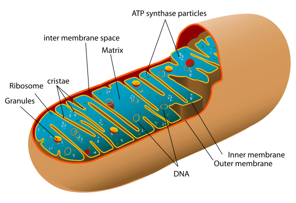
- Mitochondria are the site of production of most of the eukaryotic cell’s ATP, which provides the energy for other cellular processes.
- Plant cells contain structures not found in animal cells: chloroplasts, a cell wall. The vacuole in most plant cells is much larger than the vacuoles found in animal cells.
- The cell can be studied using a variety of experimental techniques: the electron microscope reveals the structure of the cell, cell fractionation isolates individual organelles and, with the use of radioactive isotopes in autoradiography, the passage of materials through the cell can be followed.
Thanks for reading.
REFERENCES
http://cellbiologyolm.stevegallik.org/node/74
https://en.wikipedia.org/wiki/Cell_fractionation
https://www.labce.com/spg579126_red_blood_cell_rbc_size_variation.aspx
https://en.wikibooks.org/wiki/Cell_Biology/Introduction/Cell_size
https://en.wikipedia.org/wiki/Autoradiograph
https://courses.lumenlearning.com/boundless-biology/chapter/eukaryotic-cells/
https://en.wikipedia.org/wiki/Eukaryote
https://en.wikipedia.org/wiki/Vacuole
http://lifeofplant.blogspot.com/2011/05/chloroplasts-and-other-plastids.html
https://www.ncbi.nlm.nih.gov/books/NBK9905/
https://en.wikipedia.org/wiki/Chloroplast
https://en.wikipedia.org/wiki/Symbiogenesis
Great read these types of topics, thanks for this!
I'm glad you found the post interesting, @carloserp-2000. Thanks for coming by.
Keep sharing excellent quality content;)
This post has been voted on by the SteemSTEM curation team and voting trail. It is elligible for support from @curie and @utopian-io.
If you appreciate the work we are doing, then consider supporting our witness stem.witness. Additional witness support to the curie witness and utopian-io witness would be appreciated as well.
For additional information please join us on the SteemSTEM discord and to get to know the rest of the community!
Thanks for having added @steemstem as a beneficiary to your post. This granted you a stronger support from SteemSTEM.
Thanks for having used the steemstem.io app. You got a stronger support!
Thanks.
There's a great new YouTube series called Journey to the Microcosmos that literally dives deep into the microscopic universe.
Thanks, @inertia. I'd have a look.
Hi @loveforlove!
Your post was upvoted by Utopian.io in cooperation with @steemstem - supporting knowledge, innovation and technological advancement on the Steem Blockchain.
Contribute to Open Source with utopian.io
Learn how to contribute on our website and join the new open source economy.
Want to chat? Join the Utopian Community on Discord https://discord.gg/h52nFrV