[ESP-ENG] Técnica Radiográfica de Pulmón. (Lo que debes saber) 👩🏽⚕️☢️ // Lung Radiographic Technique. (What you should know) 👩🏽⚕️☢️
Hola amigos de Hive. Como ya saben muchos, me dedico al estudio de las imágenes del cuerpo, siendo mi carrera Imagenologia y Radiofisica Médica. En el artículo de hoy les hablaré un poco del tema. En específico de una de las técnicas radiográficas más realizadas en mi campo. Se trata de las técnicas del sistema respiratorio. Específicamente de los pulmones.
Hello friends of Hive. As many of you already know, I am dedicated to the study of body imaging, my career being Medical Imaging and Radiophysics. In today's article I will tell you a little about the subject. Specifically one of the most performed radiographic techniques in my field. It is about the techniques of the respiratory system. Specifically of the lungs.
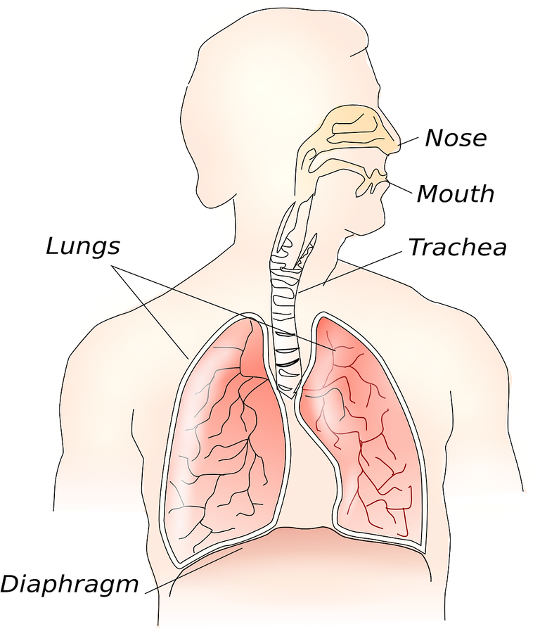
Primero que todo repasemos un poco la anatomía del sistema respiratorio:
Comenzamos con la laringe, que junto con la nariz, forman la nasofaringe, con función respiratoria. Luego la laringe y la boca, dando lugar a la orofaringe, con función digestiva además de respiratoria. En el caso de la laringe junto con la faringe su función es plenamente digestiva. Esta última va de la segunda vértebra cervical hasta la 6, donde en el borde inferior de esta comienza la tráquea, con ramificaciones a las cuales se conectan los pulmones.
First of all, let's review a little bit the anatomy of the respiratory system:
We start with the larynx, which together with the nose, form the nasopharynx, with respiratory function. Then the larynx and the mouth, giving rise to the oropharynx, with digestive as well as respiratory function. In the case of the larynx together with the pharynx, its function is fully digestive. The latter goes from the second cervical vertebra to the 6th, where the trachea begins at its lower edge, with ramifications to which the lungs are connected.
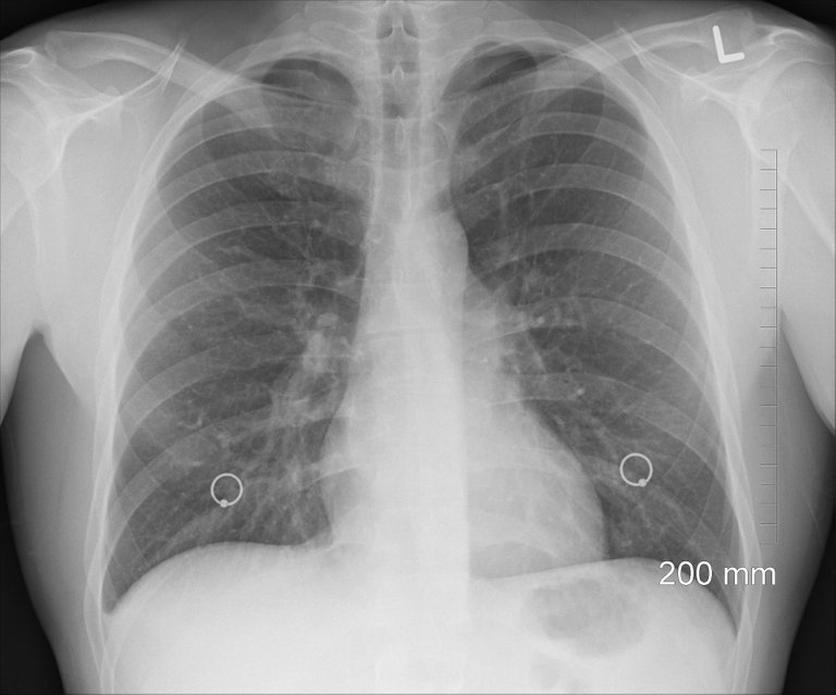
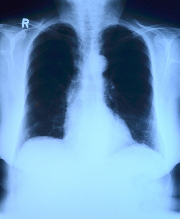
Importance of chest radiography:
The chest X-ray, is of utmost importance. Because it allows us to find bone malformations, dislocations, fractures and bone lesions.
It gives us information of the respiratory system, with the objective of observing pulmonary lesions. Also of the cardiovascular system, in pathologies of the heart and great vessels. And also of the digestive system.
Importancia de la radiografía de tórax:
La radiografía de tórax, es de suma importancia. Debido a que nos permite encontrar malformaciones óseas, luxaciones, fracturas y lesiones óseas.
Nos da información del aparato respiratorio, con el objetivo de observar lesiones pulmonares. También del sistema cardiovascular, en patologías del corazón y grandes vasos. Y además del aparato digestivo.
It is important, when performing these techniques, to take into account some details, which allow us to avoid false diagnoses, such as:
Whenever possible, perform the technique, with the patient standing or sitting, since many intra thoracic disorders (internal region of the thorax), such as pneumothorax, or width of the mediastinum, to mention a few..., are difficult to evaluate when the radiograph is performed in decubitus (patient lying down).
Also to take into account, the anatomical obtaining of the organs, as real as possible to their normal size, for this the film focus distance (distance between the patient and the focus of x-rays) should be 180 cm.
Another detail is that if it is a female patient, and the breasts are very large, as to overlap the lower part of the lung fields, the patient is asked to move them upwards and sideways.
The patient's deep inspiration is also fundamental; an X-ray performed with insufficient inspiration may simulate diseases such as pulmonary congestion or wide mediastinum.
Es importante, al realizar estás técnicas tener en cuenta algunos detalles, que nos permiten evitar falsos diagnósticos, tales como:
Siempre que sea posible, realizar la técnica, con el paciente de pie o sentado, ya que muchos trastornos intra torácicos ( región interna del tórax), como neumotórax, o anchura del mediastino, por mencionar algunos..., son de difícil evaluación cuando se realiza la radiografía en decúbito ( paciente acostado)
También a tener en cuenta, la obtención anatómica de los los órganos, lo más real a su tamaño normal, para esto la distancia foco película ( distancia entre el paciente y el foco de rayos x) debe ser de 180 cm.
Otro detalle, es si se trata de una paciente femenina, y las mamas son muy grandes, como para superponerse a la parte inferior de los campos pulmonares, se pide a la paciente que las desplace hacia arriba y hacia los lados.
Es fundamental también la inspiración profunda del paciente, una radiografía realizada con una inspiración insuficiente puede simular enfermedades tales como congestión pulmonar o mediastino ancho.
Para concluir, el estudio de los pulmones cuenta con tres técnicas fundamentales: Frontal de Pulmones con el paciente en posteroanterior; Lateral, donde se observan las regiones retroesternal y retrocardiaca; y Pancoast, la cual se utiliza en pacientes con dolor de "punta de costado", buscando derrame pleural. (Consiste en la acumulación de líquido entre los pulmones y la pared torácica, específicamente en el espacio pleural. Esto ocurre con frecuencia en las personas que desarrollan cáncer).
To conclude, the study of the lungs has three fundamental techniques: Frontal Lung with the patient in posteroanterior; Lateral, where the retrosternal and retrocardiac regions are observed; and Pancoast, which is used in patients with "side tip" pain, looking for pleural effusion. (It consists of the accumulation of fluid between the lungs and the chest wall, specifically in the pleural space. This occurs frequently in people who develop cancer).
Las causas más comunes para realizar este tipo de estudios son: bronquios primarios y secundarios inflamados, bronquitis aguda como consecuencia de gripes o referidos, enfisema pulmonar, adenocarcinoma, entre otras.
The most common causes for this type of studies are: inflamed primary and secondary bronchi, acute bronchitis as a consequence of influenza or referred, pulmonary emphysema, adenocarcinoma, among others.
Eso es todo por hoy amigos de Hive. Espero mi artículo les haya servido de apoyo en cuanto a temas de ciencias médicas se trata y lo hayan disfrutado muchísimo, principalmente los amantes de la radiología.
That's all for today friends of Hive. I hope my article has been of help to you in terms of medical sciences and that you have enjoyed it very much, especially those who love radiology.

Felicitaciones ✨✨✨
Congratulations @lorenaolivera! You have completed the following achievement on the Hive blockchain and have been rewarded with new badge(s):
Your next target is to reach 600 upvotes.
You can view your badges on your board and compare yourself to others in the Ranking
If you no longer want to receive notifications, reply to this comment with the word
STOPTo support your work, I also upvoted your post!
Check out the last post from @hivebuzz:
Su post ha sido valorado por @goya
Excelente aporte, el RX de tórax es una herramienta muy útil para complementar un diagnóstico certero.
Un RX de tórax incluso puede ayudar a salvar una vida.
Bendiciones.