Ecógrafo / Ultrasound scanner
Ecógrafo
Ultrasound scanner
Hola amigos de la colmena, después de unas vacaciones vuelvo con mucha ilusión y ganas de enseñarless un montón de equipos medicos de los que puedes aprender y tener más conocimientos, y como siempre digo "el conocimiento nunca sobra". En esta ocasión les voy a enseñar qué es un ecógrafo, cómo funciona y sus partes, así que allá vamos.
Hello my friends of the hive, after a holiday I am back with great enthusiasm and desire to show you a lot of equipment from which you can learn and have more knowledge, and as I always say "knowledge is never superfluous". This time I am going to show you what an ultrasound machine is, how it works and its parts, so here we go.

Que es la Ecografía
What is Ultrasound
La ecografía es una prueba diagnóstica que utiliza ondas sonoras de alta frecuencia para observar determinados órganos situados en el interior del cuerpo que no se pueden visualizar directamente. El uso más extendido de este estudio es en el área de la obstetricia y la ginecología debido al proceso del embarazo, pero también se utiliza en otras áreas para obtener imágenes de vital importancia para detectar y diagnosticar posibles enfermedades y anomalías en el cuerpo de un paciente.
An ultrasound is a diagnostic test that uses high-frequency sound waves to observe certain organs located inside the body that cannot be visualised directly. The most widespread use of this study is in the area of obstetrics and gynaecology due to the process of pregnancy, but it is also used in other areas to obtain images of vital importance in order to detect and diagnose possible illnesses and anomalies in a patient's body.
Que es un Ecógrafo
What is an Ultrasound Scanner
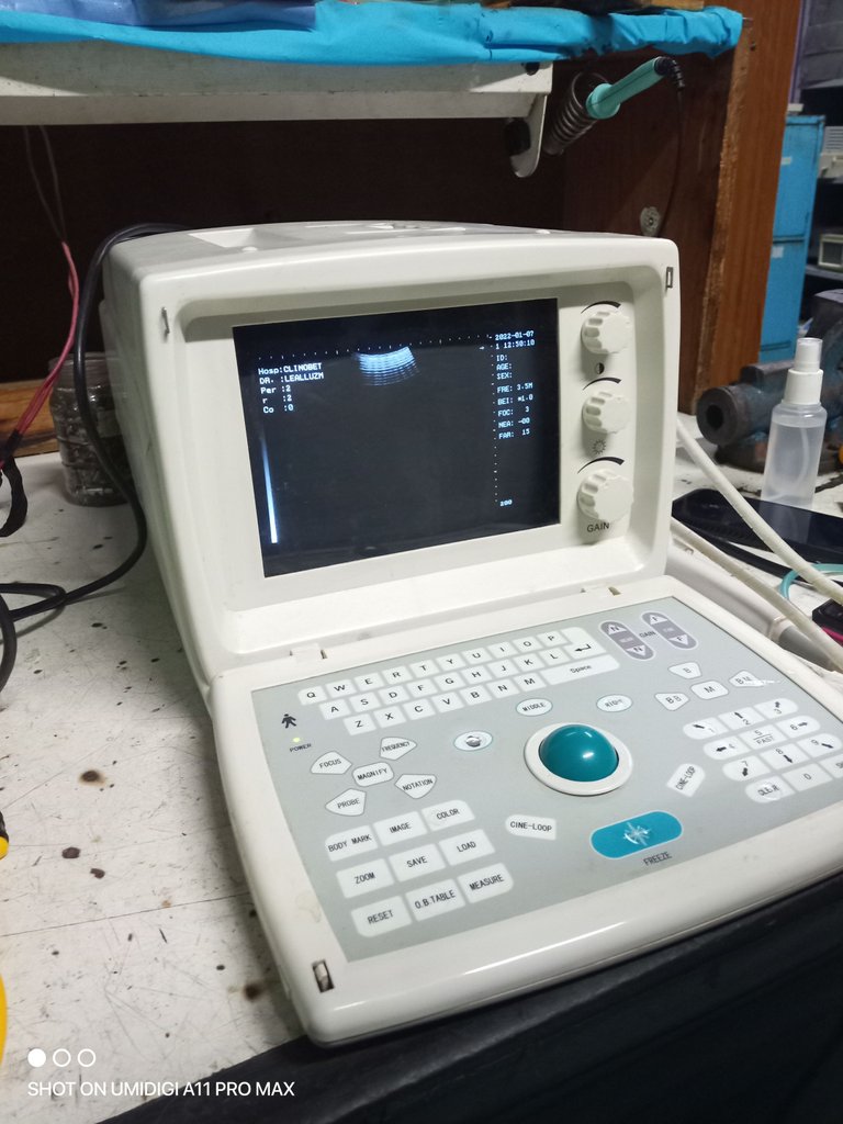
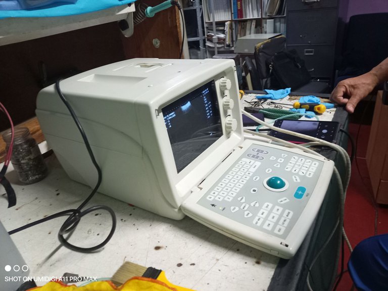
El ecógrafo es un instrumento médico que genera secuencias de imágenes del interior del cuerpo, lo que permite ver y diagnosticar posibles anomalías en los órganos y otras formaciones. También se utiliza para controlar el desarrollo del feto durante el embarazo. Los ecógrafos emiten ultrasonidos, imperceptibles para el oído humano, que penetran en el cuerpo hasta llegar a los distintos órganos que se quieren analizar. Una vez que estos ultrasonidos llegan a los órganos internos, se produce un efecto rebote que sirve para devolvernos los ultrasonidos en forma de imagen.
The ultrasound scanner is a medical instrument that generates image sequences of the inside of the body, making it possible to see and diagnose possible anomalies in the organs and other formations. It is also used to monitor the development of the foetus during pregnancy. Ultrasound scanners emit ultrasounds, imperceptible to the human ear, which penetrate the body until they reach the different organs to be analysed. Once these ultrasounds reach the internal organs, a rebound effect is produced, which serves to return the ultrasounds to us in the form of an image.
Principio de Funcionamiento
Operating Principle
Los ultrasonidos se producen mediante transductores, dispositivos formados por uno o varios cristales con propiedades piezoeléctricas. El efecto piezoeléctrico consiste en que una onda mecánica (como el sonido) provoca un cambio en la distribución de cargas eléctricas en ciertos materiales, generando un impulso eléctrico.
Ultrasound is produced by transducers, devices consisting of one or more crystals with piezoelectric properties. The piezoelectric effect is that a mechanical wave (such as sound) causes a change in the distribution of electrical charges in certain materials, generating an electrical impulse.
Se aplica una corriente eléctrica a un cristal piezoeléctrico que vibrará, según su tamaño, a una determinada frecuencia (de 2 a 10 MHz). Inmediatamente después de que el cristal genere el impulso, se pone a dormir a la espera del eco. Este ciclo se produce muchas veces con una alta velocidad. Se obtiene una mejor calidad de imagen utilizando frecuencias más altas, pero por otro lado, la penetración de las ondas es menor a frecuencias más altas. Por este motivo, se utilizan frecuencias más altas para examinar órganos más superficiales (a partir de 10 MHz) y frecuencias más bajas (entre 2 y 6 MHz) para examinar órganos más internos.
An electric current is applied to a piezoelectric crystal which will vibrate according to its size at a certain frequency (2 to 10 MHz). Immediately after the crystal generates the pulse, it goes to sleep waiting for the echo. This cycle occurs many times with a high speed. Better image quality is obtained by using higher frequencies, but on the other hand, the penetration of the waves is lower at higher frequencies. For this reason, higher frequencies are used to examine more superficial organs (from 10 MHz) and lower frequencies (between 2 and 6 MHz) are used to examine more internal organs.
Para que las ondas se propaguen correctamente, deben aplicarse a estructuras con un alto contenido de agua, por lo que no todos los órganos del cuerpo pueden explorarse con ultrasonidos, ya que algunos de ellos tienen un gran número de cavidades de aire. Los órganos aptos para la ecografía son las llamadas estructuras blandas, que son el corazón, el hígado, los riñones, el páncreas, la vesícula biliar, la vejiga, el bazo y las estructuras vasculares.
In order for the waves to propagate correctly, they need to be applied to structures with a high water content, which is why not all organs in the body can be scanned with ultrasound, as some of them have a large number of air cavities. The organs that are suitable for ultrasound scanning are called soft structures, which are the heart, liver, kidneys, pancreas, gall bladder, bladder, spleen and vascular structures.
Partes de un Ecógrafo
Parts of an Ultrasound Scanner
- Transductor
- Transducer
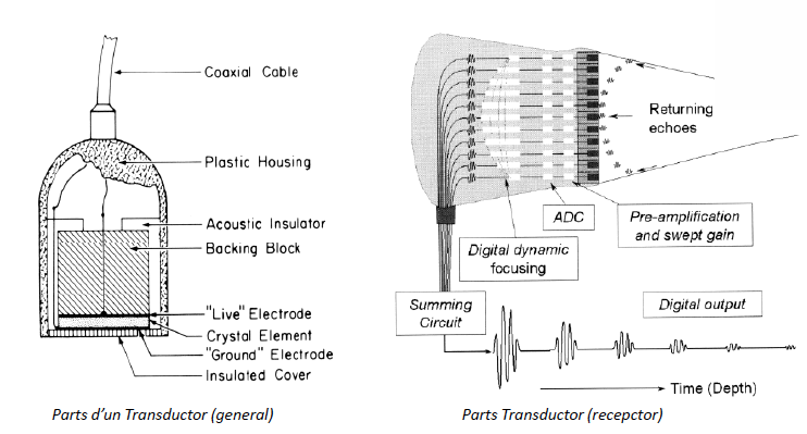
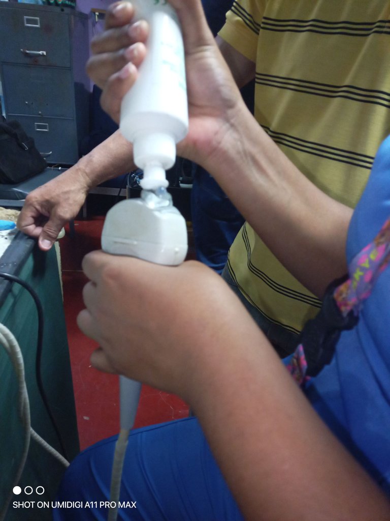
Los transductores, también conocidos como sondas, son los encargados de emitir los ultrasonidos a los órganos del cuerpo. Como ya se ha dicho, están formados por una serie de cristales llamados piezoeléctricos, cuya función es transformar la energía eléctrica en vibración o energía mecánica, y que se encuentran en el interior del transductor. Gracias a estos cristales y a la conversión de energía que realizan, la sonda envía los ultrasonidos que, al rebotar en los órganos internos, son recogidos de nuevo por los cristales piezoeléctricos y transformados de nuevo en energía eléctrica para generar las imágenes. Así pues, los transductores de ultrasonidos son una parte esencial de las imágenes por ultrasonidos.
The transducers, also known as probes, are responsible for emitting ultrasound to the organs of the body. As already mentioned, they are made up of a series of crystals called piezoelectrics, whose function is to transform electrical energy into vibration or mechanical energy, and which are found inside the transducer. Thanks to these crystals and the energy conversion they perform, the probe sends out the ultrasound, which, when bounced off the internal organs, is picked up again by the piezoelectric crystals and transformed back into electrical energy to generate the images. Thus, ultrasound transducers are an essential part of ultrasound imaging.
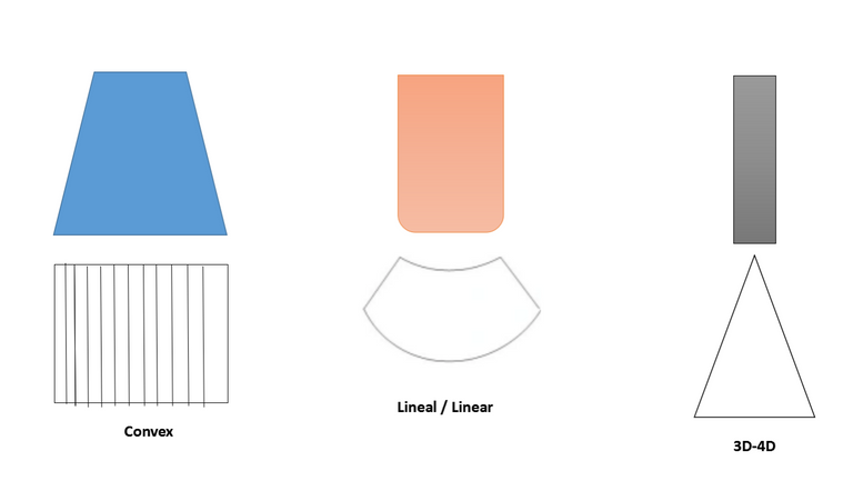
Cada ecógrafo tiene 4 tipos de transductores que son:
Each ultrasound machine has 4 types of transducers which are:
| Tipo | Descripción |
|---|---|
| Cónvex | Tienen una forma ligeramente curva. Trabajan a bajas frecuencias y tienen profundidades de hasta 30 cms. La ecografía de Abdomen y Obstetricia es con mucho su uso principal |
| Lineales | Línea de elementos recta. Pueden ampliar el campo visual gracias a su imagen trapezoidal. Frecuencias altas hasta 18 MHz. Partes blandas, músculo, estudios vasculares, ecografía ocular…tienen gran versatilidad. |
| Sectoriales | Utilizan tecnología phased array. Tienen forma cuadrada y campo visual estrecho proximalmente y muy ancho distalmente. Cardiología es su uso común. |
| 3D-4D | tienen un motor aunque también funcionan como transductores normales. Podemos variar el ángulo de barrido o hacer un barrido (3D) o hacer barridos continuos generando varios volúmenes por segundo dando lugar a la imagen 4D muy utilizada en el estudio de la ecografía prenatal. |
| Type | Description |
|---|---|
| Cónvex | They have a slightly curved shape. They work at low frequencies and have depths of up to 30 cms. Abdomen and obstetric ultrasound is by far their main use. |
| Linear | Straight line of elements. They can enlarge the field of view due to their trapezoidal image. High frequencies up to 18 MHz. Soft parts, muscle, vascular studies, ocular ultrasound...they have great versatility. |
| Sectoral | They use phased array technology. They are square in shape and have a narrow field of view proximally and very wide distally. Cardiology is their common use. |
| 3D-4D | have a motor although they work as normal transducers as well. We can vary the scanning angle or make a sweep (3D) or make continuous sweeps generating several volumes per second giving rise to the 4D image widely used in the study of prenatal ultrasound. |
- La Computadora
- The Computer
se encarga de procesar toda la información que se obtiene del transductor y la transforma en las imágenes que se muestran a través de la pantalla para que puedan ser interpretadas por el médico. Para que esto ocurra la computadora utiliza un sotfware que puede interpretar esta información. Cabe mencionar que actualmente muchos equipos nuevos traen un sistema operativo muy parecido a windows o en otros casos uno que corre en este sistema.
is responsible for processing all the information obtained from the transducer and transforming it into the images displayed on the screen so that they can be interpreted by the physician. For this to happen, the computer uses software that can interpret this information. It is worth mentioning that many new computers nowadays come with an operating system very similar to windows or in other cases one that runs on windows.
- Monitor
- Monitor
La pantalla o monitor puede variar segun el fabricante, esta pueda ser de diferentes modelos y tamaños; pero basicamente su funcion es muy sencilla trasminitar la imagen obtenida y procesada.
The screen or monitor may vary according to the manufacturer, it may be of different models and sizes; but basically its function is very simple, to display the image obtained and processed.
Aunque no forman parte de los componentes de un ecógrafo, hay dos componentes que suelen utilizarse con estos instrumentos. Uno de ellos es una impresora, con la que se pueden imprimir y tener imágenes de las ecografías realizadas en papel, y el otro es el carro para transportar el ecógrafo portátil allá donde se vaya.
Although they do not form part of the components of an ultrasound scanner, there are two components that are often used with these instruments. One of them is a printer, with which you can print and have images of the ultrasound scans performed on paper, and the other is the trolley to transport the portable ultrasound scanner wherever you go.
Fallas Frecuentes y como Identificarlas
Frequent Faults and How to Identify Them
Una de las fallas mas frecuentes en estos equipos ocurre con el transductor, ya que este parte es una de las mas delicadas y los medicos especialistas aunque tratan de tener cuidado, algunas veces y por diferentes motivos dejan caer el equipo o golpean el transductor. Como se menciona anteriormente aqui se encuentran unos cristales, estos cristales se rompen con facilidad anten golpes bruscos del equipo. Es por esto que se puede perder por completo o en parte la imagen captada por el equipo.
One of the most frequent failures in this equipment occurs with the transducer, as this part is one of the most delicate and although the medical specialists try to be careful, sometimes and for different reasons they drop the equipment or hit the transducer. As mentioned above, there are crystals in the transducer, and these crystals are easily broken when the transducer is hit by the equipment. This can result in the complete or partial loss of the image captured by the equipment.
Otra falla es que el equipo por algún problema ocasionando en la fase electrica (fluctuaciones, fallas electricas, entre otros) el equipo se apaga automaticamente y en este transcurso pierde informacion o no realiza el guardado correctamente y es por esto que al momento de realizar algun procedimiento de diagnostico el equipo no muestra una imagen en especifico.
Another fault is that due to some problem occurring in the electrical phase (fluctuations, electrical failures, among others) the equipment shuts down automatically and in this course it loses information or does not save correctly and that is why at the time of performing a diagnostic procedure the equipment does not show a specific image.
En la parte del teclado el TrackBall (bolita) tiende a no emitir la señal del moviento, eso se debe al la suciedad que se interna entre el espacio del trackball y el soporte, esta tapa los sensoceros y no capta el roce del movimiento.
On the keyboard side the TrackBall tends not to emit the movement signal, this is due to the dirt that gets between the trackball and the support, this covers the sensors and does not pick up the friction of the movement.
Espero que les haya gustado este post, no olvidés votar, compartir y seguirme para estar al día de toda la nueva información que subo sobre equipamiento médico.
I hope you liked this post, don't forget to vote, share and follow me to keep up to date with all the new information I upload about medical equipment.
El banner es de mi autoria, realizado en Picsart.
The banner is my own work, made in Picsart.
Enlaces de Interes / Links of Interest
Hola ! Me encanto tu post, esta muy bueno tu artículo ! Saludos 👍🏻🙌🏻
Muy agradecido contigo por tomar tu tiempo en leer mi post 🤗🤗
Muy buena explicación, gracias por compartirla , saludos
Al contrario gracias a ti por leer mis post ♥️🤗🤗
Primera vez que me encuentro con uno de tus post, me parece genial tu contenido
Muchas gracias por tu apoyo 🤗
Su post ha sido valorado por @ramonycajal
Gracias por su apoyo 🤗
Aprender siempre es bueno, y las explica detalladas ayudan más. Muy bien.