SENSES AND BEHAVIOUR: Structure of The Eye and Vision at Molecular Level.
The eyes are literally our window on the
world. This most sensitive and intricate of sense organs has fascinated both
scientists and poets for centuries. In this section, I will look at the basic
structure of the eye, before going on to study focusing and the correction of
defects. Finally, I will look at vision at the molecular level.
STRUCTURE OF THE HUMAN EYE
The eye is an important and delicate structure. The eyes of humans and other mammals are contained in the orbit, a bony socket of the skull. They are protected by the bone and also by a pad of fat at the rear of each eyeball that helps to cushion it from shocks. Other features of the eye are also protective: the eyebrows prevent sweat running from the forehead into the eye, the eyelashes prevent the entry of airborne particles such as flying insects, often adding to the efficiency of the eye blink reflex. Tears constantly bathe the surface of the eye, removing the surface film of dust and dirt and killing bacteria. The conjunctiva, a thin, transparent membrane which covers the cornea, also protects the eye from airborne debris.

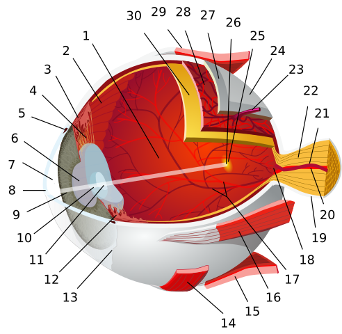
A detailed depiction of eye using a 3D medical illustration. https://www.scientificanimations.com, CC BY-SA 4.0
The human eye. The eyeball itself is a sphere about 2.5 cm in diameter. It has three basic layers: the inner retina containing the light-sensitive cells, the pigmented choroid layer and the tough outer coat, the sclera.
The main body of the eye is divided into two parts by a biconvex lens. This lens contains transparent lens fibres (long thin cells that have lost their nuclei) together with an elastic lens capsule made of glycoprotein. The lens is flatter at the front than at the back and is soft and slightly yellow. With age, it becomes flatter, yellower, harder and less elastic; it can also become cloudy. There is no blood in the lens – substances diffuse in and out of it from the surrounding fluid.
The eyeball has three cavities: the region between the cornea and the iris, the region between the iris and the lens, and the cavity that fills all the space behind the lens. The first two cavities are filled with a fluid called the aqueous humour, which is thin and watery. The third cavity, the largest (it takes up about 80 per cent of the volume of the eyeball) is filled with a thicker fluid called the vitreous humour. This transparent gel has the consistency of egg white and contains 99 per cent water and 1 per cent collagen fibres and hyaluronic acid. The vitreous humour preserves the spherical shape of the eyeball and helps to support the retina.
The retina
The retina is the light-sensitive layer at the back of the eye. It is a complex structure with a deep layer of light-sensitive cells called rods and cones, together with a middle layer of bipolar neurones which connect the rods and cones to a surface layer of ganglion cells. Fibres from the ganglion cells join up to form the optic nerve. Only the back of the retina is photosensitive. The front surface extends over the choroid and forms the inner lining of the iris and ciliary body.
The sensitive part of the retina has the area of a ten pence coin and acts as a projection screen onto which images are directed. Strangely, the nerves that come from the retina pass in front of the light-sensitive cells, but these do not interfere with vision.
Nocturnal animals such as cats have a shiny backing to the retina, a layer of cells called the tapetum. Their eyes seem to glow if a light shines into them. The tapetum reflects light back on to the receptors, making vision more effective in low-light conditions.
The choroid
The choroid contains many blood vessels. Blood supplies the cells of the eye with nutrients and oxygen and removes waste. The choroid is dark-coloured because of a high concentration of the pigment melanin in its cells. These pigmented layers prevent internal reflection of light rays and so prevent us seeing a confused and blurred image.
At the front of the eye, the choroid expands around the edge of the lens to form the ciliary body. The smooth muscles in the ciliary body alter the shape of the lens. The lining of the ciliary body secretes aqueous humour, the fluid that fills the front of the eye.
The iris is also an extension of the choroid, partially covering the lens and leaving a round opening in the centre, the pupil. Its function is to control the amount of light entering the eye. The iris contains radial and circular muscles that can alter pupil size. Iris muscles are controlled by the autonomic nervous system.
The sclera
The sclera forms the ‘white’ of the eye. It is a tough external coat mainly a collagen fibres. Six exterior muscles attached to the sclera enable the eye to look up, down and side-to-side. The central one-sixth of the sclera is colourless and transparent, forming a clear window, the cornea.
HOW THE EYE WORKS
The process of seeing is complex and involves five different stages:
- Light enters the eye.
- An image is focused on the retina.
- Energy in the light that makes up the image is transduced into an electrical signal.
- Nerve impulses carry information about the image into the brain.
- The brain decodes the information and perceives the image.
Let’s look at each of the stages in more detail.
Light enters the eye
Light enters through the cornea and then passes through the aqueous humour, the pupil, the lens and the vitreous humour, before it reaches the retina. To operate well in conditions of different light intensity, the eye must control how much light reaches the retina. It does this by changing the diameter of the pupil. In bright light, the pupil constricts (gets smaller) to prevent overstimulation of the retina and the perception of ‘dazzle’. In dim light, the pupil dilates (gets wider), letting in more light.
The size of the pupil is controlled by the muscles in the iris. These are themselves controlled by the autonomic nervous system and so pupil adjustment is a reflex response, not under the conscious control of the brain.
Focusing light on the retina
If you do not focus a camera correctly, the picture you take is blurred. Similarly, the eye must focus light to produce a high-resolution image on the retina. The eye focuses an image by refracting or bending light using the cornea and the lens, forming an upside down or inverted image on the retina.
Because of the composition and curvature of the cornea, most of the refraction of light occurs in this structure. The lens is important in the fine focusing of light onto the retina. The cornea is fixed but the lens is adjustable, allowing accommodation, a reflex that makes the eye focus on objects that are at different distances from the eye.
Accommodation
Generally, when we open our eyes in the morning, they are not focused on nearby objects. At rest, the ciliary muscles have relaxed, pulling the lens flat. In this state, we focus on distant objects.
When we focus on an object that is only a metre or so away, the ciliary muscles of the eye contract involuntarily. This reduces the tension on the ligaments that hold the lens in place, and the lens becomes ‘fatter’. The focal length changes, and the image is focused. When we then look at an object much further away, the ciliary muscles relax, again automatically. The tension on the ligaments supporting the lens increases, and the lens becomes thinner’, allowing our vision to accommodate.
The two eyes converge to point to the same object.. Wikimedia, CC BY-SA 3.0
PROBLEMS WITH THE LENS SYSTEM
In some individuals the lens and cornea do not focus correctly. If you are short-sighted, a condition known as myopia. You focus light from an object in front of the retina. This produces a blurred image. Short-sightedness can result from an elongated eyeball or a thickened lens. It is corrected by using a diverging or concave lens.
If you are long-sighted you are said to have hypermetropia, and you focus light behind the retina. Again, objects appear blurred. Long-sightedness results from a shortened eyeball or a lens that is too thin, and is corrected using a converging or convex lens.
A more complicated visual defect is astigmatism. In astigmatism, the surface of the cornea is irregular, the object appears blurred because some of the light rays are focused and others are not (this is similar to the distortion produced by a wavy pane of glass). Astigmatism is corrected using a cylindrical lens that bends light rays in one plane only.
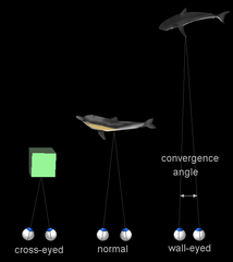
BINOCULAR VISION
Humans and some other mammals, notably primates and predators such as cats, have binocular vision. We have eyes at the front of our head, so the field of vision seen by each eye is similar but not identical. Normally, the brain is able to compute the incoming signals so that we see only one image.
Binocular vision gives us a big advantage. When two sets of signals are decoded by the brain, we get the impression of distance and depth and of objects being three-dimensional. This stereoscopic vision allows animals to judge distance and depth accurately. It is easy to see why primates need binocular vision; when living in the trees, any animal that cannot judge distance is likely to fall sooner rather than later.
Predators often have their two eyes placed centrally to maximize this overlap of retinal images. It gives them excellent stereoscopic vision to judge distances accurately and locate their prey. Prey species, on the other hand, tend to locate their eyes to the side of the head. They have reduced stereoscopic vision but an all-round vision that allows them to spot nearby predators, even if they try to sneak up from behind.
VISION AT THE MOLECULAR LEVEL
When light reaches the back of the retina it is focused on to the photoreceptors, cells that are specialised to detect light. These cells contain pigment molecules that are bleached by light. Bleaching of the pigment results in a generator potential and, when the threshold is reached, action potentials are created and nerve impulses begin to travel towards the brain.
The two types of photoreceptor in the human eye are the rods and cones.
Photoreception in rod cells
Rods contain a reddish-purple compound called rhodopsin, or visual purple. This consists of two joined compounds – opsin, a protein, and retinal, a light-absorbing compound derived from vitamin A. It is important to know what happens to rhodopsin in the dark and when it is exposed to light.
In the dark, sodium ions flow from the surrounding fluid into the outer segment of the rod cell via non-specific cation channels. The sodium ions then diffuse along the rod cell and into the inner segment, where they are actively pumped back out of the cell. The overall movement of sodium ions produces a slight depolarisation of the whole rod cell membrane. The potential difference across the membrane is about -40 mV, compared with the normal -70 mV resting potential. This slight depolarisation brings about continual release of a neurotransmitter, thought to be glutamate, which crosses the synapse and prevents bipolar cells from depolarising. Bipolar cells are the ‘on’ or ‘off’ cells that connect rod and cone cells to nerve cells in the optic nerve. The overall result is that in the dark we don’t see anything because no signals are coming from the rod or cone cells into the brain.
When light falls on the rhodopsin molecule, it breaks down into retinal and opsin. The opsin activates a series of reactions, which close the cation channels. This stops the movement of sodium into the outer segment of the rod cell but the inner segment continues to pump sodium out. The inside of the cell becomes more negative and the release of glutamate stops. No glutamate means there is nothing to prevent depolarisation of the bipolar cell and an action potential is generated and transmitted down the optic nerve. These nerve impulses pass to the brain for decoding and we see an image. In the absence of further light stimulation, the retinal molecule goes back to its original shape and then recombines with opsin to form rhodopsin. It is worth remembering that this resynthesis process takes time.
Rhodopsin is very sensitive to light and so rods can function effectively in dim light. In bright light, our rhodopsin is broken down quicker than be re-formed, so that most of it is bleached for most of the time. In this state, we are said to be light adapted. If we enter a dark room from a brightly lit one we cannot see anything because much of our rhodopsin is bleached. However, as we resynthesise the pigment and become dark adapted, things become clearer and we are able to see in dim light, even if it is only in shades of grey.
So as not to make this post too lengthy, in my next post, I shall discuss about the photoreception in cone cells, visual acuity and the transmission of nerve impulses to the brain.
Thanks for reading.
REFERENCES
https://www.msdmanuals.com/home/eye-disorders/biology-of-the-eyes/structure-and-function-of-the-eyes
https://www.britannica.com/science/human-eye
https://www.thoughtco.com/how-the-human-eye-works-4155646
https://en.wikipedia.org/wiki/Human_eye
https://en.wikipedia.org/wiki/Retina
https://www.healthline.com/human-body-maps/retina
https://en.wikipedia.org/wiki/Tapetum_lucidum
https://en.wikipedia.org/wiki/Choroid
https://en.wikipedia.org/wiki/Sclera
https://www.livescience.com/3919-human-eye-works.html
https://www.umkelloggeye.org/conditions-treatments/anatomy-eye
https://www.aoa.org/patients-and-public/resources-for-teachers/how-your-eyes-work
https://www.stanfordchildrens.org/en/topic/default?id=normal-vision-90-P02094
https://www.nkcf.org/about-keratoconus/how-the-human-eye-works/
https://www.sciencelearn.org.nz/resources/50-how-the-eye-focuses-light
https://www.visionaware.org/info/your-eye-condition/eye-health/anatomy-of-the-eye/125
https://en.wikipedia.org/wiki/Accommodation_(eye)
According to the Bible, The Mark of a True Witness: No More, No Less
Watch the Video below to know the Answer...
(Sorry for sending this comment. We are not looking for our self profit, our intentions is to preach the words of God in any means possible.)
Comment what you understand of our Youtube Video to receive our full votes. We have 30,000 #SteemPower. It's our little way to Thank you, our beloved friend.
Check our Discord Chat
Join our Official Community: https://steemit.com/created/hive-182074
We are so blessed to be granted such marvels. Thanks for reminding us.
This post has been voted on by the SteemSTEM curation team and voting trail. It is elligible for support from @curie and @minnowbooster.
If you appreciate the work we are doing, then consider supporting our witness @stem.witness. Additional witness support to the curie witness would be appreciated as well.
For additional information please join us on the SteemSTEM discord and to get to know the rest of the community!
Thanks for having used the steemstem.io app and included @steemstem in the list of beneficiaries of this post. This granted you a stronger support from SteemSTEM.