Wonders of being extra HOT (Animal tissue slide for Immunofluorescence/Cell Biology)
Hello Guys,
I am sharing one of my latest experience with you all which might be helpful for
budding life science/biology researchers. I work on Colorectal cancer ...and carry
out clinical studies too. Whoossshhhh...I know a lot to do. My interest is to
understand the role of few proteins in contributing tumor progression. As a cancer
biologists, we all know that KI-67 (antigen protein) is a nuclear protein which is
associated with any cellular proliferation. Hence, used as Cancer cell marker
protein.
Now, here it is what happened. So I was following my usual protocol of
immunofluorescence staining of animal tissue slide, and failed three
times…(OMG). But this time, I thought of being lazy...and heated the slide for a
longer duration for antigen retrieval step (and played some games during that time,
I know that's wrong), used old antibody to tag. Next day, I came and did secondary antibody addition and went for imaging. I was very sure this time also it's going to
be a waste of time as well as reagents (still I went on doing, because I had a very
small ray of hope).
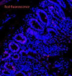
And guess what, thats was the best staining I ever did, saw beautiful images. I am
sharing my happiness (see the image, sorry little blurred though). So now I was wondering which magic step
it was? Starting from paraffin deparaffinization to dehydration in alcohol (=
wanted so much after 3 failures), then rehydration. Later comes antigen retrieval
step, where I became lazy (the magic step), so instead of heating 3 times...I did 4
times..so that I can relax more. After analysing everything step by step, and I found
that it was the heating step. I was like…..WOW..even my protein wants to be
HOT.
Even after being in research for 8 years, I still wonder which step or idea can
work when and where!
P.S. I still don't know whether it was an idea or my laziness.
(If anyone interested I can share my protocol for Ki-67 IF staining)
Hope it was fun and informative too while reading this.
Love Ruby
Posted using Partiko Android
Congratulations @excelsureacademy! You have completed the following achievement on the Steem blockchain and have been rewarded with new badge(s) :
You can view your badges on your Steem Board and compare to others on the Steem Ranking
If you no longer want to receive notifications, reply to this comment with the word
STOPVote for @Steemitboard as a witness to get one more award and increased upvotes!
I can relate to that joy when being lazy make things work better. Guess I will give it a shot too for staining ki67.
Anyhow, few questions came to my mind when reading your post. Why did you need antigen retrieval for Ki67. Was it an antibody thing or was it because of fixation you used? Were these Oct samples fixed with PFA or were these paraffin embedded tissue sections?
Also am I looking at the normal non-cancerous colon section here? Is this the basal level of ki67 that we usually find in those glands there. They are glands right? It's kind of interesting.
Anyway, thanks for sharing this image and the post. Hope to read more from you.
Those were paraffin embedded tissue sections. And yes that was from normal tissue section. The section was taken from colon.
Posted using Partiko Android