Anatomy of eye
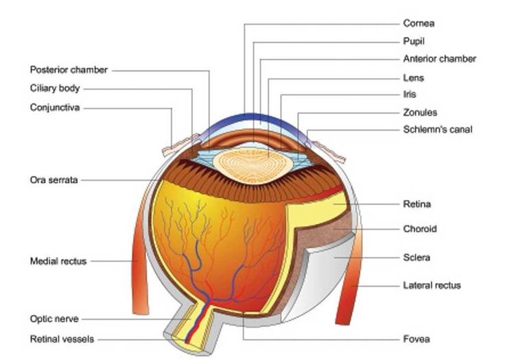
THE EYEBALL Each eyeball (Fig. 1.1) is a cystic structure kept distended by the pressure inside it. Although, generally referred to as a globe, the eyeball is not a sphere but an ablate spheroid. The central point on the maximal convexities of the anterior and posterior curvatures of the eyeball is called the anterior and posterior pole, respectively. The equator of the eyeball lies at the mid plane between the two poles (Fig.1.2). Dimensions of an adult eyeball Anteroposterior diameter Horizontal diameter Vertical diameter Circumference Volume Weight 24 mm 23.5 mm 23 mm 75 mm 6.5 ml 7 gm Coats of the eyeball The eyeball comprises three coats: outer (fibrous coat), middle (vascular coat) and inner (nervous coat). 1. Fibrous coat. It is a dense strong wall which protects the intraocular contents. Anterior 1/6th of this fibrous coat is transparent and is called cornea. Posterior 5/6th opaque part is called sclera. Cornea is set into sclera like a watch glass. Junction of the cornea and sclera is called limbus. Conjunctiva is firmly attached at the limbus. 2. Vascular coat (uveal tissue). It supplies nutrition to the various structures of the eyeball. It consists of three parts which from anterior to posterior are : iris, ciliary body and choroid. 3. Nervous coat (retina). It is concerned with visual functions.
Hi! I am a robot. I just upvoted you! I found similar content that readers might be interested in:
http://www.doctoralerts.com/eye-anatomy/
You seem to be using older or Legacy version of eSteem!
Please check if you have newest version to get most out of eSteem, Android, iOS mobile app. For desktop Windows, Mac, Linux Surfer app!
Learn more: https://esteem.app
Join our discord: https://discord.me/esteem