A very brief history of virology with Veeru
Scienceblocks: Hello Veeru, how are you?
Veeru: I am good. Why are you here again? You still think your life is a lie.
Scienceblocks: No! Your last interview has gathered a couple of interesting questions. Thought that I could ask you to answer them.
Veeru: Why do you humans have so many questions? Why can't just get along with a good virus and rest in peace?
Scienceblocks: Don't act smart I see what you did there, not cool.
Veeru: Sorry, always wanted to try it out. Anyhow, what are your questions?
Scienceblocks: There are two. One is regarding Kary Mullis and another one is about the legacy of your kind. I will reserve the question about Kary Mullis for the next post. What I want to know today is about how and when you guys slipped and got caught by humans? Basically, I want you to answer this question was asked by - @mmykel - "What year was the first viral infection detected?"
Veeru: Well! Acidically...
Scienceblocks: Don't even!
Veeru: OK! OK!
But this is a difficult question to answer. First virus infection must have been detected long before humans evolved a language. We were there, even when you did not even have a word called "virus". That is a different thing that your kind used to think all plagues and diseases are caused by evil spirits. Guess what! It was us all along (viruses, bacteria, and even those huge-ass protozoan that causes malaria). So you detected the infections long-long ago, in a galaxy not so far away. You just didn't know about it.
Scienceblocks: Oh boy! Smartypants! Stop about my kind and tell me when did your kind slip. When was the first time humans figured out that some disease is caused by the virus?
Veeru: Haha! You guys struggled pretty bad. Let me tell you a story of what all transpired. And then I will leave it up to you to make that call.
Viruses have an interesting history. The term virus itself originates from a Latin word meaning - poisonous secretion 1. The term was used in medical science to describe secretions from ulcers or pustules. Medical science then started using the term for infectious secretions. It took time. But eventually, you humans figured out that there were us, these nanometer-sized particles in those secretions.
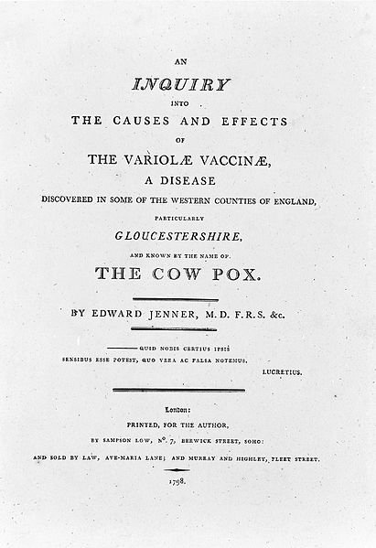
Image from Wellcome Images | CC BY 4.0
Scienceblocks: And yet, without knowing about you, my kind used the secretions from diseases caused by you to create vaccines. Didn't we now!
When Edward Jenner, published his work in 1799, about smallpox vaccination (using cowpox virus which was derived from pustules of infected cows); he titled his work - "An inquiry into the causes and effects of the variolae vaccinae"2.
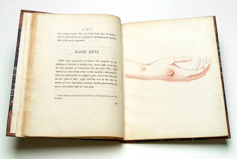
Veeru: Yes! Back then, members of my clan were still undiscovered. But, what he was referring to by the term "variolae vaccinae" was pustulate secretion from Vacca - the cow (the term which later evolved into the modern word vaccine). Nevertheless, what he was dealing with was indeed members of my clan. In fact, some of the most complicated and dangerous members if you ask me.
Scienceblocks: So it was that smallpox virus that gave you away?
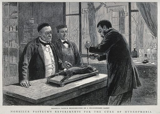
Louis Pasteur (left) watching over a colleague working with rabbit and rabies.
Wood engraving, 1885. Created 21 November 1885. Rabies. Rabbits as laboratory animals. Louis Pasteur (1822–1895). Work ID: jxdzwjnq. from Wellcome Images | CC BY 4.0
Veeru: Oh no no no!
As the legends from the times of my great grandfather say - the first us who gave us away was perhaps the rabies virus in Pasteur's lab in 1887. But, we almost got away because Pasteur thought the causative agent was some sub-microscopic microbe. It was invisible under the microscope. He probably tried to culture us like bacteria, which must have led to further disappointment. But, he cultured us in spinal cords of rabbits (that was smart!) probably because no one wanted to work with the dogs. This led to the development of rabies in rabbits. He later figured out a way to decrease virulence. He did so by drying the sections of infected spinal cords in the flask. He then used it to incubate rabbits, who got vaccinated without developing the disease.
Then in 1885, a 9-year-old child came to him who was bitten by a rabid dog. Pasteur was unsure, but the death of the child was inevitable. He injected the child with spinal cord suspension from the infected rabbits and saved his life 3.
But alas! Despite all the mounting evidence, the dogma around living microbes and bacteria haunted the scientists of that time. And they couldn't imagine the existence of something like a virus. It was not until 1962 that they took the first image of this Rabies virus.
Scienceblocks: Lucky you!
Veeru: Not for long. The idea of invisible microbe got a lot of people interested.
In 1892, a guy named Dimitrii Ivanovsky figured that the causative agent of tobacco mosaic disease in the plants can pass through filter pores designed to stop bacteria. However, he insisted that the pathogen is probably a very small bacteria or a soluble bacterial toxin that can pass through these pores 4. Even Loeffler and Frosch passed pathogen of foot and mouth disease of cattle through a filter in 1898. Making it the first animal virus to be filtered. But being students of Koch, they couldn't get over the 'one bacterium-one disease' dogma 5. Even they thought that we are some sort of very small bacteria. Lol.
Then, Walter Reed and his collogue, in 1901, became the first people to show that hemorrhagic yellow fever was spread by mosquitoes. They also demonstrated that the causative agent is filterable. Making them the first to isolate a human filterable virus. Now, I don't know what they believed about us. But then, it needed a lot more than a filter to prove that we were different.
Scienceblocks: So they came close. I see. But were blinded by Dogma and preconceived notions. So when did the bubble burst?
Veeru: Yes.
Now we don't have eyes. But legends say that in 1898 there was this very handsome guy - Martinus Beijerinck. Though he was eccentric and not very social, but was smart af! He confirmed the findings of Dimitrii Ivanovsky about tobacco mosaic disease. Furthermore, he figured out that we are different from bacteria. He figured that we depend on host cells to multiply. He thought that we must be some sort of soluble germ. He called us infectious living liquid 6.
Phew! He was damn close. Closer than anyone else came. He was also the first to use the term 'virus' as we know it today. But thanks to the dogma about pathogens being bacteria and the lack of adequate imaging techniques back then. The war of ideas raged on about us. Who are we - are we just a tiny bacteria, living liquid, or something else altogether. It boggled the mind of every scientist who came across a disease that could pass through filters.
In fact, that is what they started calling us - filterable viruses. In 1927, T.M. Rivers wrote an account on the "filterable viruses and knowledge gained by that point." He wrote -
In all fields of work, times come when one must stop and take
thought. New facts, new ideas, and new suggestions alter lines
of endeavor in every field of research. We are here today for the purpose of taking thought concerning the knowledge of the so called filterable viruses and the diseases caused by them.
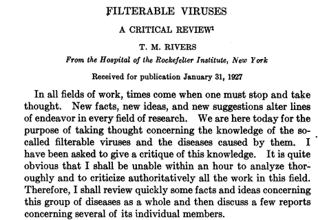
Screenshot of article by T.M. Rivers
IIn summary, in his essay 7 he pointed out certain properties of viruses that became consensus about the nature of viruses. The so-called "filterable viruses" were thought to have certain properties -
- Filterability - Unlike bacteria, they can pass through filter pores.
- Invisibility - Can't be seen with a microscope.
- Non-cultivability - They can't be cultivated in absence of living cells.
- They don't breathe - no evidence for the existence of respiration was found in viruses, not in contact with living cells.
Scienceblocks: But those are all negatives. You can't claim you found something because you have negative results.
Veeru: Damn! Yes.
But, around 1930, they created even finer filters and proved that viruses were indeed particulate matter, which can be animated only inside the host 8. Then in 1935, Wendell Meredith Stanley finally crystallized the tobacco mosaic virus. This was soon followed by x-ray distraction methods which for the first time revealed the nature of the particle 9. However, during this time, the Dogma was that viruses were infectious proteins. Based on the existing bias in the field, Stanley concluded the same with his data - TMV is an infectious protein 10. But, this idea was challenged. Later on, Stanley and his postdoc acknowledged that viruses are made of protein and RNA. Now, this was interesting. For one, it was before Watson and Crick have published the structure of DNA. Second, recently Griffth has proposed that DNA is the genetic material that makes genes. So what was the purpose of RNA? It would take them some time to figure out how viruses use RNA as genetic material, but that is a topic for another day.
Then, in 1939, the first image of TMV was taken in an electron microscope11. This is for the first time someone was looking at the shadow of these thread-like viruses on the fluorescent screen of the electron microscope.
Scienceblocks: Wow! Such a journey. So I would say it is hard to decide which viral infection should get the status of the first discovered virus. Was it Rabies virus, or was it plant virus TMV. They all had their contributions as a mole to your kind and achievement to mine. Many discoveries led to the concept of the virus we know today. I think it would be safe to assume that TMV was the first snitch.
Veeru: Oh! Talking of moles and snitches. I think the one I hate the most is the bacteriophage. It literally offered us to you guys on a plate. I mean it is what infects bacteria. I mean, it was so obvious. It's just that you guys took time. I think you should know about them too.
Scienceblocks: Oh really! What is the story of bacteriophage then?
Veeru: Well!
A submicroscopic beast in bacterial cultures has been a pain in the ass. It all started in 1896 when Ernest Hanbury Hankin observed that water from the river Ganga in India killed the bacteria that caused cholera 12, 13.
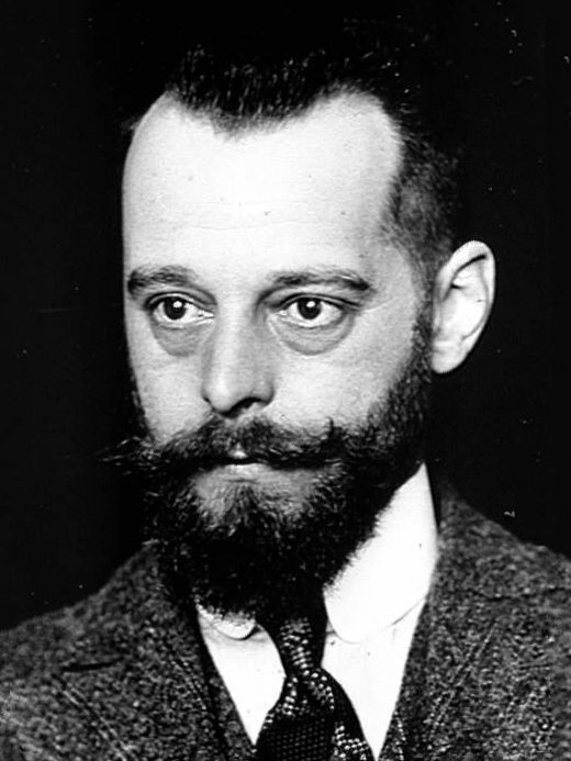
Portrait de Félix d'Hérelle, vers 1905 by Service photo Institut Pasteur - Photothèque | Public Domain
Then in 1917, Félix d'Herelle discovered that there is something in nature that can pass through filters, just like TMV did in 1898 and killed the bacteria in those agar plates. 14. He claimed that the antibacterial activity is due to an organism called bacteriophage. Which translates to bacteria eater15. Though he later became hesitant about his claims. They would learn later that it is a virus that instead uses what bacteria eat and then burst open the baterial cell to release itself. Hence, technically it is an obligate parasite and not a literal bacteria eater.
Nevertheless, just like TMV, the argument continued on what really is this bacteriophage? Is it a virus? Or, is it just a protein (an enzyme) that can digest bacteria? And yet again, in the 1930s, it was shown that these were virus particles. And they were finally imaged in 1940, and the argument was settled after all.
Damn! Those electron microscopes. You guys would have been in the total dark without them.
Scienceblocks: We have come a long way even from electron microscopes now.
Rosalind Franklin, who took the X-ray diffraction picture of DNA, also worked out more details of the structure of the TMV 16. Later, on cryo-electron microscopy was developed and even cleaner pictures of viruses were taken. And now with single electron cryo-EM, the resolution that we can achieve is mind-blowing 17.
Veeru: I mean, hats off to your species, man!
Scienceblocks: Anyhow, I guess that will be all for today. I will get back to you about the second question on Kary Mullis on the weekend.
Veeru: Oh boy! I have deal with you again on the weekend!
Scienceblocks:As if you have a job to do in this lab flask. Will you rather prefer to get bombarded with an electron beam?
Veeru: I guess, I will see you on weekend then. Caio!

#STEMsocial powered by Hive
Video by @gtg
- ‘Virus’: The Spread of a Latin Term for Poison
- An inquiry into the causes and effects of the variolae vaccinae: a disease discovered in some of the western counties of England, particularly Gloucestershire, and known by the name of the cow pox
3.Historical Perspectives A Centennial Celebration: Pasteur and the Modern Era of Immunization - Dmitri Josifovich Ivanovski (1864-1920)
- Constructing scientific culture in the 5G era: Historical lessons from the first discovery of a virus
- Beijerinck's Work on Tobacco Mosaic Virus: Historical Context and Legacy
- FILTERABLE VIRUSES. A CRITICAL REVIEW. T. M. RIVERS
- The isolation and properties of crystalline tobacco mosaic virus
- Wendell M. Stanley. Facts
- History of Virus Research in the Twentieth Century: The Problem of Conceptual Continuity
- Die Sichtbarmachung yon pflanzlichem Virus
- The bactericidal action of the waters of the Jamuna and Ganges rivers on Cholera microbes.
- Bacteriophage prehistory
- On an invisible microbe antagonistic to dysentery bacilli.
- bacteriophage (n.)
- After the double helix: Rosalind Franklin's research on Tobacco mosaic virus
- Seeing the target of vaccines with your eyes - Capturing highest resolution image of a virus (Zika virus)
You can reach out to me on discord if you are in stemsocial discord. Or you can even send me a DM. My discord handle is the same as my hive - @scienceblocks. You can also ping me on my telegram handle: @UncertainHeisenberg. Or follow me on Twitter: @scienceblocks1
Do ask if you have any questions. If I find your question very interesting I can ask veeru to explain it in a post, like this one.
Adding @mmykel as a beneficiary, since this post is the result of that question. Also adding @mycrimsonhues, for letting me use her artwork.
Kary Mullis ? I had no idea who this person was and look over the Internet.. Now I understand the interest (and my nose feels it ;) ). I am looking forward to read your next post about him, curious about what you want to say.
I really enjoyed reading this post, especially how the differences between viruses and bacteria emerged (and actually stayed hidden for so long). Really really cool (well, if this word can be used in this context ;) )!
I usually write my questions and comments all during the course of my reading. When you mentioned that viruses were invisible and cannot be seen with microscopes, I was really puzzled and assumed that this was the case in the 19th century (plus the first half of the 20th). I had however troubles to conceive this was still the case today, as we have good techniques to unravel the nano-world. And then you killed my question when you mentioned electronic microscopes. This being said, is there any more recent observations with very modern microscopes such as those used every day in nanoscience research?
I think your nose feels it right. 😁 And yes advanced microscopes are now used to produce super high res images of the virus. Years back I wrote a post in steemstem about using single particle Cryo-electron microscope to image Zika virus with resolution on Armstrong range. It was based on this cell paper, if you want to look at the images. I got a chance to prepare sample for cryo-EM back in 2020 when one of the drug we were testing appeared to cause aggregation of virus particles. Now that wasnt some super high res image, but I could clearly see the spikes of CoV2 protuding out of a sphere of about 100nm.
Other than cryo-EM Xray-diffraction is common method to get atomic structure of viruses. But that work best with viruses you can crystalize.
Even the popular nanoscience tool - atomic force microscope is also used. Check out some AFM inages of the virus in this paper. What I find cool about AFM is that it can show you viruses entering in or budding out of the cells.
Ahaha I totally miss this post! The image you shared is great (and the comments to the post are hilarious; yeah the good old times). Also, thanks for the papers. They have really nice images (I only checked out the pics; I didn't read the text).
Cheers!
again a creative and well-written article, it was fun to read :D. I'm happy that they discovered the actual viruses at the end and the genetic stuff after WW2.
I have so many questions 😅. If viruses are so old (as old as life), how does a virus in a single cell organism work? If you are a nano- or pico-trophist you eat viruses for breakfast I guess?
It works pretty much the same way as it does in a single cell of a multicellular organism. For example the phage particle attaches to a bacterial cell and then injects the genome inside using its tail. The genome can either get integrated into the bacterial genome and replicate with bacteria. Or it can make virus proteins and copies of its genome. In the later case it will burst open the bacterial cell and released the assembled phage particles out in the open.
Image by Thomas Splettstoesser | CC BY-SA 3.0
That being said, bacteria also have tools to fight back and degrade the phage genome that enters the bacterial cells. I mean that is how we got the current CRISPR technology. Its always an evolutionary arms race there. Here is a post titled - Journey from adaptive immunity in bacteria(CRISPR) to saving Man's best friend(Dog). which I wrote sometime back regarding how CRISPR works. You may find it interesting.
Cominh to your second question on virus eaters. There aren't many examples of it. Its mostly like virus infects the cells and if virus fails the cells degrades its constituents and probably use it for something. Now there was some ideas floating around that some protists may eat viruses in labs, but there have not been any hard evidence for it. However recently, Brown et al., in 2020 claimed that some protists esp picozoans might eat viruses. They think their eating aparatus is too small to consume bacteria, but large enough to consume viruses of 150nm and use them as carbon source. I mean they do need more evidence. But for now I can do a more detailed journal club on why this is an interesting paper if it interests people.
Let me know if you have any further questions. 🙂
Hi there,
you are using an image and language here that refers to visible things from the animal world we know. We know "eating and being eaten", "proboscises and appendages that dive into something from the insect world".
But when we talk about atomic/sub-atomic processes, this language is inappropriate because we cannot see what these particles are doing.
We can only translate it into a language that serves the human imagination through the instruments that make something visible with the help of chemistry, rays and other substances.
In this respect, I consider such interpretations as have been captured here in the picture to be rather the expression of an artist or designer, but not of what is happening there on the atomic or molecular level.
This form of language is highly suggestive; our habit of understanding something through visual images is strongly served here.
Imaging or imaging processes that serve as mediators between the invisible and visible worlds are inventions that now go far into the interpretive realm because they do not allow what can be seen with the eye or with the microscope to be seen without further intervention. But this raises some concerns about accuracy.
The more you rely on a combination of manipulation of states through the use of these techniques, the more justified it is to ask whether it is not the manipulation itself that has influenced the very outcome of what is made visible. I rarely see this question asked.
The layman will think of an electron microscope as a better machine that reinforces the old image of "I am looking through a magnifying glass". However, it is impossible to depict a real-time image of such small particles in the form shown here. Therefore, it is and remains a linguistic interpretation.
Correctly one should say "it was made for me to see" instead of saying "I saw it".
I can partly understand the great fascination that such imaging techniques using physics and chemistry provide. They create the impression of extensive control over even those processes that are not accessible to our normal senses. They are meant to provide a sense of security precisely where we are no longer sure that what we are looking at is what we are (not) seeing. If that makes sense to you?
The samples that are examined for such imaging, have they in turn already been affected at a hierarchical level higher up by chemicals or aggregation state change processes? I assume so. So what is left of the original nasal or throat swab? Is the original still there?
Thanks for your contribution to the STEMsocial community. Feel free to join us on discord to get to know the rest of us!
Please consider delegating to the @stemsocial account (85% of the curation rewards are returned).
Thanks for including @stemsocial as a beneficiary, which gives you stronger support.
I have a newfound admiration for Pasteur after reading this.