CLASS OF REPTILIA, AVES(BIRD), AND MAMMALS. INTRODUCTION TO CELLS
The phylum of chordata is a wide phylum that have so much of classes subordinating.
CLASS REPTILIA
The reptiles are mainly terrestrial animals that, for many millions of years, were the dominant group of animals on the Earth.
Their characteristics of physical attributes is that they have dry skin with scales and their gas exchange takes place exclusively in the lungs. They have developed internal fertilisation and they lay eggs on land in a leathery shell. Some reptiles even keep the head within their body and give birth to fully developed young.
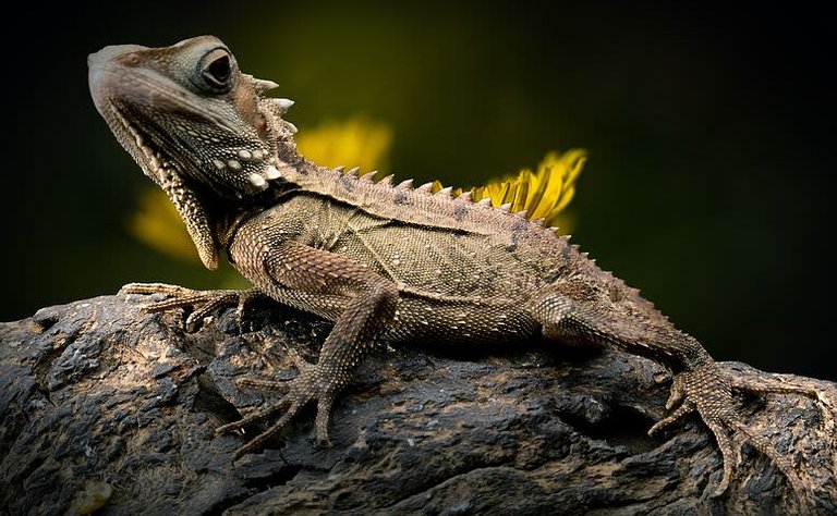
Reptiles are found in many areas of the world. Reptiles have no external ears. Reptiles are ectothermic, meaning that they rely on heat from their environment to regulate their body temperature. Examples include snakes, crocodiles and lizards such as the red- headed rock lizard.
CLASS AVES(BIRD)
Class Aves comprises the birds. Birds are endothermic, which means they use heat produced by their own metabolism to regulate their body temperature. Birds have feathers over most of the body, and scales on their legs. The forelimbs are adapted as wings which most birds use to fly.

The sternum (breastbone) is enlarged into a big keel shape. This provides an attachment of the wing muscles, particularly in those birds which fly. Birds have light skeletons that make it easier for them to fly. The Jaws are toothless and are covered by a horny beak. Birds reproduce using well- developed eggs with a hard shell. In most species, the parent birds spend a lot of time and effort raising their young. Examples include domestic fowl, the golden-crowned woodpecker, hornbills and Kingfishers.
CLASS MAMMALIA
Mammals differ from other Chordates in a number of ways. A true mammal produces milk for it's young in mammary glands, has a high internal body temperature and regulated its own body temperature. Most mammals sweat to help control their body temperature. A mammal has hair on the skin and external ears. Most mammals also produce live young that have developed for a time within the body of the mother in a structure called the uterus.
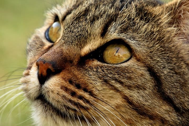
The mammals range in size from tiny shrews to elephants and whales. The way that control their body temperature and reproduce inside their bodies had made it possible for them to live almost everywhere on the earth.
SUB-DIVISION OF MAMMALS
Mammals are classified according to how their young are produced. There are three sub-classes of mammals.
(1). Egg laying mammals: lay eggs, for example duckbilled platypus
(2). Marsupials- produce immature young that are nourished by milk in pouch, for example kangaroo, koala bear, opossum
(3). Higher mammals: produce fully developed young that are nourished by milk from the mammary glands, for example rats, cows, elephants, cats, monkeys and humans.
INTRODUCTION TO CELLS
Everything that we know about the structure of cells is due to the development of the microscope. Microscope were developed more than 300 years ago. This led to the development of the cell theory. This theory stated that the cell is the basic unit in all living organisms.
The best light microscope will magnify up to around 1500 times. Light microscopes have given us a lot of information about the structure of cells. However, in the last 50 years we have also been able to use electron microscopes. An electron microscope can give a magnification of around 500000 technology had made it possible to gain lot more information about cells.
THE GENERAL STRUCTURE OF A CELL
Organisms are made up of cells. Some contain only a single cell. Other organisms contain an enormous number of cells working together.
There are some basic structures that are found in all cells, from single- celled organisms to the cells in complex animal and plant cells. For example, almost all cells have a nucleus, a surface cell membrane, mitochondria, ribosomes and cytoplasm.
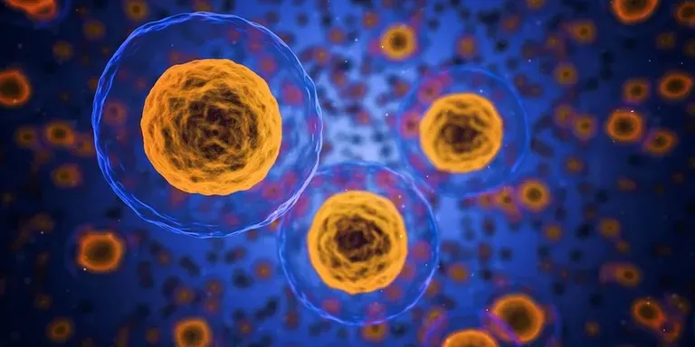
Other features are often seen in plant cells, particularly from the green parts of the plants, but not in animal cells. This has led scientist to develop a picture of the basic structure of an unspecialized animal cell and an unspecialized green plant cell.
Although not many cells are quite this simple, the idea of unspecialized animal and plant cells gives you a very useful model with which to compare other, more specialized cells.
STRUCTURES AND FUNCTIONS IN AN UNSPECIALIZED ANIMAL CELL
You can see the structures that are common to all cells in an unspecialized animal cell. Many of these structures (or organelles) contains enzymes and chemicals to carry out specialized jobs within the cell.
(1). The nucleus controls all the activities of the cell. It also contains the instructions for making new cells or new organisms in the form of long threads known as chromosomes. This is the genetic material.
(2). The cytoplasm is a liquid gel in which most of the chemicals reactions needed for life take place. About 70 percent of the cytoplasm of a cell is actually water. As a result, your body is around 70 percent of the cytoplasm of a cell is actually water. As a result, your body is around 70 percent water as well! The cytoplasm contains all the other organelles of the cell.
(3). The cell surface membrane forms a barrier like a very thin skin around the outside of the cell. The membrane controls the passage of substances such as carbon dioxide, oxygen and water into and out of the cell. Because it lets some substances through, but not others, it is known as a selectively permeable membrane.
(4). Mitochondria (singular: mitochondrion) are the powerhouse of the cell. They carry out most of the reactions of respiration, where energy is released from food in a form that your cell can. Cells that need a lot of energy such as muscle cells and secreting cells, contain many mitochondria.
(5). Ribosomes are found on stacks of membranes in your cells.( These stacks are known as the endoplasmic reticulum). Ribosomes are vital for protein synthesis.
(6). Animal cells sometimes contain a vacuole a fluid filled space but they are never permanent.
Most cells can only be seen under the microscope but some are very large indeed. The biggest single cell is an unfertilised ostrich egg which is around 18 cm long, 14 cm wide and weighs about 1.5 kg.
HOW TO USE THE MICROSCOPE TO LOOK AT ANIMAL CELLS
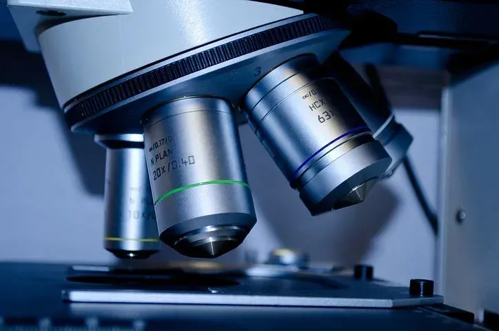
(1). Set up your microscope with the lowest power lens(the smallest lens) in place.
(2). Clip the prepared slide into place on the stage using the stage clips. Position the specimen over the hole in the stage.
(3). If your microscope has a built-in lamp, switch it on. If it has a mirror, adjust the angle of the mirror until the specimen is illuminated.
(4). Now look through the eyepiece lens and adjust the Iris diaphragm until the light is bright. But doesn't dazzle you. The illuminated area you can see us known as the Field if view.
(5). Looking at your microscope from the side(not through the eye peice lens) and using the coarse focusing knob, move the objective lens down slowly so it is as possible to the slide without touching it.
(6). Now look through the eye peice lens again. Turn the coarse focusing knob very gently in the opposite direction to move the objective lens away from the slide. Do this while you are looking through the eye peice lens and the specimen will gradually appear in focus. Once you can see the specimen clearly, use the fine focusing knob to get the focus as sharp as you can.
(7). You may find that if you now shut the Iris diaphragm down further, so that the hole for the light to pass through get smaller, you will see the specimen better (the contrast is greater).
(8/. To use the higher magnifications, rotate the nosepiece so that the next lens clicks into place. Do not adjust the focusing knobs at this point as the specimen should still be in focus and, with the coarse focusing knob in particular, it is very easy to break the slide. If you do need to adjust the focus, use the fine focusing knob only with higher magnifications. Take great care to avoid letting the lens touch the slide.
(9). Human check cells and simple epithelial cells are very similar to the diagram of an unspecialized animal cell. Remember you will not see ribosomes or mitochondria under normal light microscopes.
CONCLUSION
Now that we have been able to look at the introduction of cells, in.my next post, I will be going deeper in talking more about cells
REFERENCES
. https://en.m.wikipedia.org/wiki/Reptile
. https://byjus.com/biology/aves/
. https://en.m.wikipedia.org/wiki/Mammal
. https://www.sparknotes.com/biology/cellstructure/intro/summary/
Congratulations @mandate! You have completed the following achievement on the Hive blockchain and have been rewarded with new badge(s) :
You can view your badges on your board And compare to others on the Ranking
If you no longer want to receive notifications, reply to this comment with the word
STOPTo support your work, I also upvoted your post!
Support the HiveBuzz project. Vote for our proposal!
Tweets
https://mobile.twitter.com/adenijiadeshin7/status/1274348875021778944
#posh
Thanks for your contribution to the STEMsocial community. Feel free to join us on discord to get to know the rest of us!
Please consider supporting our funding proposal, approving our witness (@stem.witness) or delegating to the @stemsocial account (for some ROI).
Thanks for using the STEMsocial app and including @stemsocial as a beneficiary, which give you stronger support.