NERVOUS SYSTEMS: The Brain and its parts.
The human brain weighs about 1.5 kilograms, is 85 per cent water and has the consistency of thick blancmange. It is, however, the most complex material known. It copes with a huge amount of information from the various senses, deciding what is important and what can be ignored. It stores thousands and thousands of memories for decades, and can sort them into chronological order. It allows us to control the complex functions of the body, while at the same time allowing us to maintain posture, read, write and talk. Can we make a computer to do all of this? Not a chance.
The brain is the organ that makes humans human. We owe our success to this remarkable mass of nerves which takes over 20 years to mature, and which allows us to make tools and use language to an extent that far surpasses our nearest relative, the chimpanzee.
The brain of a human makes up about one-fiftieth of the body’s mass. Its delicate tissues are protected by the skull or cranium and by the cranial meninges, membranes that are continuous with the spinal meninges. Cerebrospinal fluid bathes the outside of the brain and fills the chambers – the ventricles. Twelve pairs of cranial nerves innervate (supply nerves to) various regions of the head. The human brain is thought to contain ten thousand million (1010) neurons. Each neuron may be in contact with a thousand other cells, providing an immense number of different communication routes.
The areas of the brain
The brain receives a vast number of impulses from receptors both inside and outside the body. It maintains basic involuntary body, ‘housekeeping’ functions such as heart rate, breathing rate and temperature control. It also co-ordinates the semi-automatic muscular actions of the body, such as swallowing, and it initiates and controls voluntary activities such as walking and running. The human brain is the site of higher mental functions such as reasoning, emotion and personality.
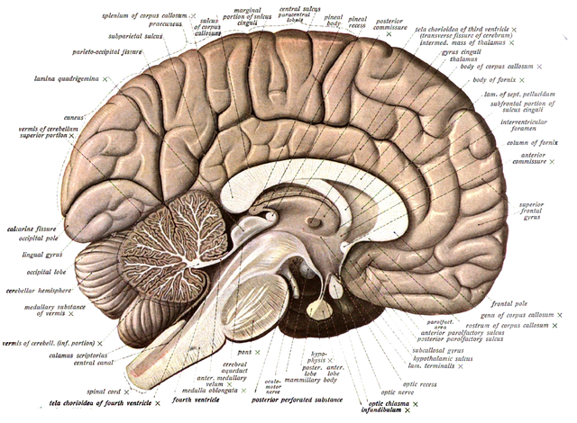
Like all vertebrate brains, the human brain consists of three parts, a hindbrain, midbrain and forebrain. However, our brains have evolved and enlarged to such an extent that the basic three-part pattern is only noticeable in the early stages of development of the embryo.
The hindbrain has three distinct structures. The medulla oblonga is a swollen portion at the bottom of the brain stem that houses vital centres controlling heart rate, breathing and blood supply. The cerebellum controls body movement and maintains balance, and the reticular activating system (a collection of neurones in the centre of the brain stem) filters incoming stimuli and controls wakefulness and sleep. The brain stem is the medulla oblongata plus the pons plus the midbrain.
As in other animals, the midbrain in humans links the forebrain to the hindbrain. Our emotions, which are located in the forebrain, can affect basic functions of the hindbrain such as control of blood vessel diameter, heart rate and sweating. When we are worried about something – exam results for example – the forebrain interprets this as stress and brings about the release of adrenaline, a hormone that prepares the body for action. The forebrain has two main parts, the cerebrum and a region containing the thalamus and the hypothalamus.
Hypothalamus
The hypothalamus is a key area. It receives a huge amount of internal and external sensory information and acts as a co-ordinating centre between the nervous system and the endocrine (hormonal) system. The hypothalamus is responsible for sensations such as hunger and thirst. It also helps control the autonomic nervous system, since it regulates body temperature and the balance of water and salts in the blood. It is linked directly to the pituitary gland by means of blood vessels and nerves.
Generally, the hypothalamus controls the release of hormones from the pituitary gland, including the antidiuretic hormone (which controls water reabsorption in the kidneys), growth hormone and some reproductive hormones. The thalamus directs sensory information from the sense organs to the correct part of the cerebral cortex.
Cerebrum
The cerebrum is made up of two large cerebral hemispheres. They have a thin outer layer, the cortex, which is thrown into many folds with fissures (grooves) in between. The cortex is the surface layer of the hemispheres, and most of our conscious thought takes place here. The Cerebral cortex carries out the ‘higher’ mental activities of reasoning, and is regarded as the site of personality and emotion and a sense of ‘self’. This is what sets humans apart from other species.
Different areas of the cerebral hemispheres are associated with different sensory and motor functions. The cerebrum of mammals is very large compared with the forebrain of other vertebrates and is certainly the dominant feature of the human brain. Fold in the surface of the cerebrum increase the surface area for centres of control, where incoming nerve impulses are interpreted, or integrated, in the light of information already stored in the brain.
Remember the following terms: Neuron – a nerve cell. Stimulus – a change in an organism’s environment (internal or external) that can be detected by receptor cells. Receptor – a specialized cell that detects a stimulus and initiates a nerve impulse. Sensory neurone – a nerve cell that carries impulses from the receptor into the central nervous system. Central nervous system (CNS) – the brain and spinal cord. The CNS processes the incoming information and decides on a response often based on previous experience. Motor neurones – nerve cells that carry impulses from the CNS to the effectors. Effectors – the organs that bring about the response, usually muscles or glands.
Overall, the cerebral cortex can be divided into three areas according to their function:
- The sensory areas receive sensory information.
- The information is processed and interpreted in association areas, which decide on an appropriate response.
- Responses are initiated in the motor areas and modified by the cerebellum.
In this section I will look at each of these areas in turn.
The sensory areas of the cerebrum
The sensory areas of the brain receive incoming impulses from the vast number of receptors that provide us with information about the world outside. The external receptors might tell us that it is cold, dark, raining and that the skin is getting wet. Your brain has to decide what to do. There are many receptors in the skin, but, as you know, some parts of the body are more sensitive than others. The lips, tongue and fingertips have more receptors per unit area of skin than anywhere else.
The association areas of the cerebrum
The association areas interpret the sensory information and make decisions in the light of previous experience, i.e. our memory. If you are cold and wet, you may see a friend’s house and your visual association areas will compare it with the images of houses stored away. Once you have recognized it, impulses pass to your frontal lobes, which then make a conscious decision to knock at the door.
The way information from the eyes is processed gives us a particularly impressive example of brain function. The millions of rods and cones in the retinas of both eyes generate a stream of impulses that pass down the optic nerves to the visual cortex at the back of the brain. The different information from the two eyes is processed in the visual association area into a single image in which we have a perception of depth as well as colour and detail.
Speech and language
We take it for granted that we can listen to someone speaking our own language and understand what they say, but have you ever wondered how this is possible?
When we hear speech, the organ of Corti in the ear converts the sounds to nerve impulses that travel to the primary auditory cortex in the cerebellum of both hemispheres of the brain. The two sides recognize different information in the signals. Both then send signals to an area called Wernicke’s area in the left hemisphere. This is responsible for processing the sounds of speech. Wernicke’s area is therefore the speech association area.
People who have damage in this area of the brain cannot understand someone speaking to them in their own language, even though they can hear perfectly. When they speak, it sounds OK in terms of rhythm but the sounds don’t make any sense. This is a condition known as receptive aphasia.
In the normal brain, after the signals have been processed in Wernicke’s area, the information is then passed to another area called Broca’s area through a bundle of nerves called the arcuate fasciculus. Broca’s area further processes the signals and transmits processed information to nearby motor-related areas. These connect with the parts of the face, jaw and throat and are responsible for speech production. So, Broca’s area is the speech motor centre.
People with damage to Broca’s area can understand what others say to them but they can’t actually speak. This condition is known as expressive asphasia. Broca did a lot of his work with a patient with a damaged Broca’s area who could only say the word ‘tan’.
So, in summary, Wernicke’s area lets you understand what someone else says, Broca s area lets you work out your reply.
The motor areas of the cerebrum
The motor areas generate the impulses that cause the muscles to contract. This simple statement hides the fact that the process is incredibly complex and requires such a degree of computation that we are unlikely to be able to make a real lifelike ‘android’ robots for a long time to come.
Muscular contraction is not a matter of all or nothing. If it were, muscles would be either relaxed or fully contracted. Movement, if possible at all, would be very jerky. However, we can move our muscles with a great deal of precision, creating just the right tension for the action we are performing.
This is made possible by the process of proprioception, in which the brain receives constant feedback about the degree of contraction of the muscles and the position of the joints in relation to each other. Vital information also comes from the inner ear about our position with respect to gravity, direction of movement, acceleration and deceleration. The process of movement control and postural maintenance is a matter of countless fine adjustments to alter the tension in the muscles. Interestingly, the left side of the body is controlled by the right motor area, and vice versa.
There are three key components to the process of moving:
- selection of a motor programme;
- initiation of a motor programme – the muscles contract;
- Modification of movement in the light of feedback.
Given the complexity of the task, It is not surprising that several different areas of the brain are involved in the control of movement.
The process starts with the basal ganglia, a collection of structures lying deep in the forebrain. These are of fundamental importance because they select and initiate the motor programme.
In the image showing the pathway taken by motor impulses before they reach the muscles; from the basal ganglia impulses pass to the motor cortex.
The motor cortex contains two major regions, the primary motor cortex and the supplementary motor cortex. The primary motor cortex contains neurons that send impulses to skeletal muscles along nerve fibres passing down the brain stem and spinal cord.
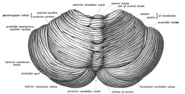
The cerebellum is a complex structure lying on the posterior surface of the brain stem. Its function is to co-ordinate ongoing movements, thus producing smooth flowing movements. The cerebellum uses information from proprioceptors to decide whether the body’s actions are going to plan or need modifying.
REFERENCES
- https://www.webmd.com/brain/picture-of-the-brain
- https://www.britannica.com/science/brain
- https://en.wikipedia.org/wiki/Human_brain
- https://www.hopkinsmedicine.org/health/conditions-and-diseases/anatomy-of-the-brain
- https://courses.lumenlearning.com/wmopen-psychology/chapter/outcome-parts-of-the-brain/
- https://mayfieldclinic.com/pe-anatbrain.htm
- https://www.health24.com/Mental-Health/Brain/Anatomy-of-the-brain/Brain-areas-and-their-functions-20120721#:~:text=The%20brain%20can%20be%20divided,Hippocampas%20and%20the%20Mid%2D%20brain.
- https://en.wikipedia.org/wiki/Cerebrum
- https://www.britannica.com/science/cerebrum
- https://en.wikipedia.org/wiki/Primary_sensory_areas
- https://courses.lumenlearning.com/boundless-ap/chapter/functional-systems-of-the-cerebral-cortex/
- https://www.neuroscientificallychallenged.com/glossary/association-areas#:~:text=Association%20areas%3A,between%20sensory%20and%20motor%20areas.
- https://en.wikipedia.org/wiki/Wernicke%27s_area
- https://www.verywellmind.com/wernickes-area-2796017#:~:text=Wernicke's%20area%20is%20the%20region,to%20the%20production%20of%20speech.
- https://en.wikipedia.org/wiki/Organ_of_Corti
- https://www.understood.org/en/learning-thinking-differences/child-learning-disabilities/communication-disorders/difference-between-speech-impairment-and-language-disorder#:~:text=Speech%20refers%20to%20the%20actual,sounds%20that%20make%20up%20language.
- https://courses.lumenlearning.com/boundless-ap/chapter/functional-systems-of-the-cerebral-cortex/
- https://en.wikipedia.org/wiki/Motor_cortex
- https://nba.uth.tmc.edu/neuroscience/m/s3/chapter03.html#:~:text=The%20motor%20cortex%20comprises%20three,motor%20area%20
!discovery 30
@tipu curate
Upvoted 👌 (Mana: 21/28)
Thanks for your contribution to the STEMsocial community. Feel free to join us on discord to get to know the rest of us!
Please consider supporting our funding proposal, approving our witness (@stem.witness) or delegating to the @stemsocial account (for some ROI).
Thanks for using the STEMsocial app and including @stemsocial as a beneficiary, which give you stronger support.