Neurology Explained - The Vestibulocochlear nerve (Cranial Nerve VIII) || The Cochlear (Auditory) Pathway
After taking time to explain the cochlear, the vestibule, and the semicircular canal, I will be looking into the vestibulocochlear Nerve which is the 8th Cranial Nerve. In case you missed out on the previous cranial nerve posts I made in the past, the link to each of the cranial nerves are in the box below.
Neurology Explained - The Oculomotor nerve (The Cranial Nerve 3)
Neurology Explained - The Trochlear Nerve (The Fourth Cranial Nerve)
Neurology Explained - The Trigeminal Nerve (The Fifth Cranial Nerve)
Neurology Explained - The Abducens Nerve (The Sixth Cranial Nerve)
Neurology Explained - The Facial Nerve (The Seventh Cranial Nerve)
The ear performs two basic functions which are hearing and balancing. The vestibulocochlear nerve has a major role in those functions, as the vestibulocochlear nerve has both the cochlear nerve and the vestibular nerve combined. The Vestibulocochlear nerve has the Auditory System as well as the vestibular system.. I will be picking it one after the other, so let's kick off with the hearing/auditory aspect of the cranial nerve VIII.
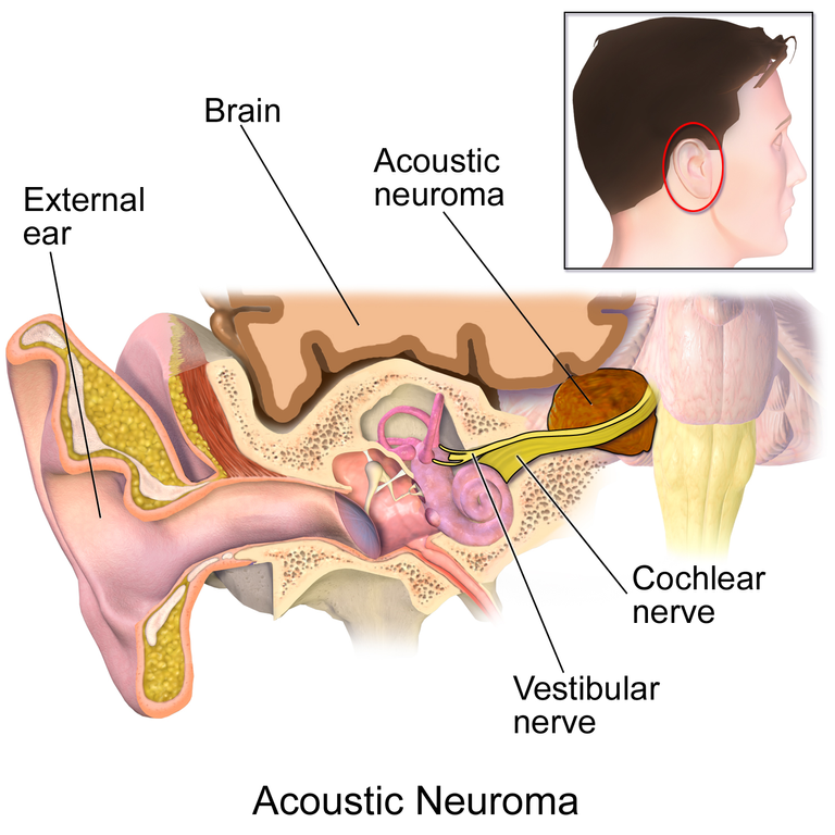
Remember in my post, I talked about the process of hearing starting from the Ear auricle (Pinnae), you should read my previous post on The Anatomy, and Physiology of the Ear, the get a better understanding. The ear auricle helps to send the sound waves through a sigmoid shaped tube that attaches to the pinna, known as the external acoustic meatus, in other to get the sound into the ear. After which the external acoustic meatus takes the sound the tympanic membrane (ear drum), which is a connective tissue structure that divides the external ear from the middle ear.. The sound wave causes a vibration the shakes the bones of the middle ear. These bones are the Malleus, Incus and Stapes. The ear ossicle stapes vibrates and tap the oval window which seperates the middle ear from the inner ear.. Once the Stapes hit the oval window, it causes mechanical energy to be transferred in form of fluid-filled vibration through superior cochlea duct known as the Scala Vestibuli which causes vibration to the basilar membrane in the Scala media, as well as causing the hair cell (stereocilia to beat towards the kinocelium),then the vibration goes to the scala duct , which hits the round window.. When the Stereocillia beats towards the kinocilium in the scala media, it release glutamate (neurotransmitter) to the affarent nerve, which transports the neurotransmitter to the pseudounipolar neuron ganglion, known as the Spiral ganglion.,.
The Spiral gangion has a peripheral process as well as a central process. The peripheral process goes to the organ of corti where the hair cells are, while the central process goes to the central nervous system.. Spiral ganglia central processes form to become the cochlear fibres (cochlear branch) in the cranial nerve VIII.,,.
The Cochlear nerve fibres goes through the internal acoustic meatus to reach the Pontine-medulla (pontomedulla) junction. Other nerves that go through the internal acoustic meatus are the vestibular nerve (to form the vestibulocochlear nerve), facial nerve (CN VII), and the labyrinthine artery. At the pontine medulla junction, it goes to the Cochlea nuclei which are of two types, the Ventral cochlear nuclei, and the Dorsal Cochlear nuclei. . The Ventral cochlea nuclei are sub-divided into the anteroventral cochlear nuclei (AVCN), and posteroventral cochlear nuclei(PVCN),. The majority of the fibers of the Dorsal Cochlear Nucleus ascend to the Nucleus of the contralateral lateral lemniscus in the midbrain, the other fibres ascend in the ipsilateral lateral lemniscus..
The posteroventral cochlear nuclei (PVCN) made up of octopus cells, move across to the nucleus of the contralateral lateral lemniscus via its fiber known as the intermediate acoustic stria, the anteroventral cochlear nuclei (AVCN) axons known as the ventral acoustic stria or the trapezoid body, go to the contralateral lateral lemniscus and synapses to the ipsilateral lateral lemniscus. Fibers from the nucleus of the contralateral lateral lemniscus, and fibers from ipsilateral lateral lemniscus move together to become the lateral leminiscus from both nucleus. The lateral lemniscus move to inferior colliculus.. From the inferior colliculus, a part of the fibers branch to become the Tectospinal tract, a descending motor pathway responsible for auditory reflexes, while another branch goes to the Brachium of inferior colliculus (inferior brachium), which carries the auditory fibers to the medial geniculate nucleus in the thalamus. The sound stimulus from the medial geniculate nucleus goes to the primary auditory cortex, then to the superior temporal gyrus, then to the Wernicke's area for speech comprehension.. To respond, there are connections known as Arcuate fasiculus between the Brocas area responsible for speech and the Wernicke's area for comprehension, .
From the Superior Olivary Nucleus, the olivocochlear bundle releases acetylcholine to the hair cells cochlear, to reduce the basilar membrane, thereby decreasing the action potentials released, thereby helping to reduce hearing and its range, and it causes the reticular formation on the auditory receptor to activate the cranial nerve 5 and 7. The cranial nerve 5 activates the tensor tympani to pull the malleus from vibrating and dampening the sound stimulus (read more on the cranial nerve 5 post). The cranial nerve 7 activates the stapedius to pull the stapes from tapping the round window (read more about this in the cranial nerve 7)..
Conclusion
The vestibulocochlear nerve as I said at the beginning, starts with the outer part of hear as sounds go into the hear. They reach the cochlear, whenre the hair cells in the cochlear triggers the spiral ganglion whose axons serves both peripheral and central processes. The central processes go to the pontine-medulla junction through the internal acoustic meatus. The nerve fibers reaches the cochlear nuclei. The Dorsal Cochlear Nucleus reaches the contralateral lateral lemniscus, the Ventral Cochlear nucleus made up of two nuclei, also send fibers to the contralateral lateral lemniscus and the ipsilateral lateral lemniscus. The nerves from this nuclei move to the inferior colliculus, then to the primary auditory cortex where certain bodies which includes the Superior Temporal Gyrus, ansd the Wernicke's area mediates sound stimulus and speech comprehension.
Image Reference
Image 1 || Wikimedia Commons ||Acoustic Neuroma
Congratulations @eni-ola! You have completed the following achievement on the Hive blockchain and have been rewarded with new badge(s):
Your next target is to reach 45000 upvotes.
You can view your badges on your board and compare yourself to others in the Ranking
If you no longer want to receive notifications, reply to this comment with the word
STOPCheck out the last post from @hivebuzz:
Support the HiveBuzz project. Vote for our proposal!
Thanks for your contribution to the STEMsocial community. Feel free to join us on discord to get to know the rest of us!
Please consider delegating to the @stemsocial account (85% of the curation rewards are returned).
Thanks for including @stemsocial as a beneficiary, which gives you stronger support.