Neurology Explained || The Components of the Midbrain
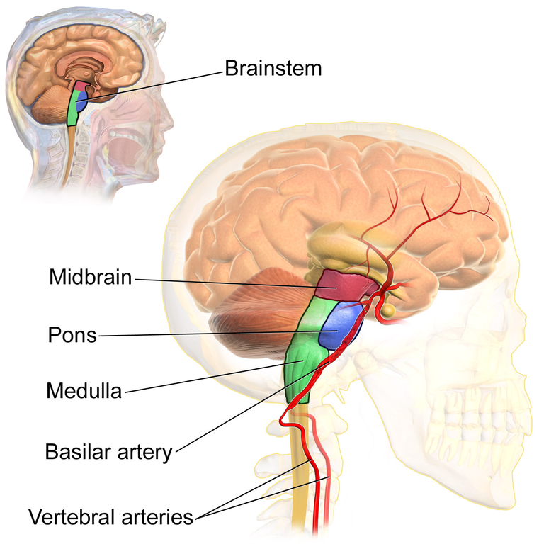
In my previous post, I went over the overview of the anatomy of the Midbrain. In that post, I explained the inferior colliculus which is at the dorsal part of the midbrain and the superior colliculus which is at the ventral part of the midbrain, and I mentioned the components of both colliculi. In both colliculi there are several components which i mentioned in my previous post, and in this post, I will be writing about this components individually and their function. Let's begin;
- Lateral Lemniscus
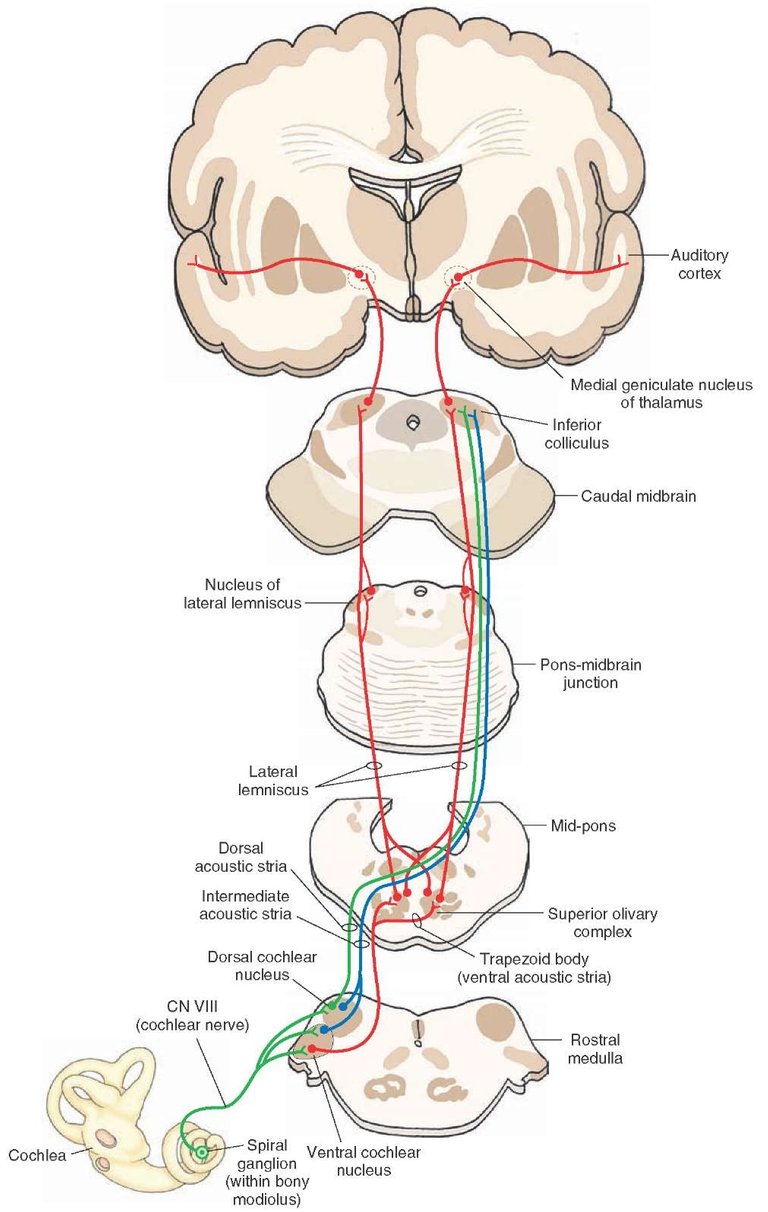
In my post on the vestibulocochlear nerve, do you remember the spiral organ of corti? The vestibulocochlear nerve recieves innervation from the spiral organ of corti. The sensation goes to through the vestibulocochlear nerve to the pontomedulla junction where it gets to the cochlear nuclei where they synapse and cross, moving up through the lateral lemniscus which eventually synapse at the inferior colliculus which sends auditory information to the medial geniculate nucleus which is outside the thalamus which will be sent to the temporal lobe. In simple terms, the lateral leminiscus is responsible for carrying the sensory auditory pathway to the inferior colliculus which then sends it to the medial geniculate nucleus.,,. When audtory sounds come from the inferior colliculus to the superior colliculus, the Tectospinal pathway is activated, which sends the auditory signals to the motor neurons which supplies the muscles that supply the head and the neck.,
- Crus Cerebri
Upper motor neurons which are situated at different lobe in the cortex send their fibers through the internal capsule then to the midbrain in a descending fiber where it gets to the level of the Pons where they synapes with the Pontine nuclei and then move downwands where they get to the lateral columns then towards the anterior gray horn in the spinal cord where the lower motor neurons are stimulated causing muscle contraction. The fiber from the upper motor neuron to the anterior gray horn is known as the Corticospinal fibers. While they move towards spinal they can branch in the brainstem to stimulate nuclei which are called corticobulbar fibers, while the fibers that synapse at the pons are the Corticopontine fibers. At the pons where the fibers in the corticopontine crosses through the middle cerebellar peduncles where it connects through the cerebellum.,,,.
- Substantia nigra
The Substantia nigra through the nigrostriatal pathway sends fibers to connect to the striatum which controls the cordination of motor neurons. The fibers from the Substantia nigra connects to the Lentiform nucleus which is made up of the putamen, the globus politis external, and the globus pallidus internal which are cornnected to the thalamus. The substantia nigra is involved in two pathways, which are the direct and the indirect pathways. The pathway stimulates motor movement in the cortex, while the indirect pathway inhibits motor movement in the cortex. The substantia nigra releases dopamine to the neurons involved in the direct pathway and indirect pathway. A lesion in this area is responsible for parkinson.,,.
- Decussation of the Superior cerebellar peduncles
To explain this, let me start with the cerebellum where the Deep cerebellar nuclei which includes the dentate, the globose, and emboli nuclei send fibers known as the cerebello rubral fibers to the controlateral red nucleus or they may go directly to the thalamus known as cerebellothalamic fibers while the fibers from the red nucleus go to the thalamus, then to the motor cortex. The red necleus would send some fibers downwards to the inferior olives which send the fibers back to the cerebellum, to ensure the cordination of motor movement.,
- Lemnicus
Actually I have discussed the lateral lemnicus above, but there are more beyond the lateral lemnicus. There is the medial lemniscus,spinal lemniscus, and the trigeminal lemniscus. To explain medial lemniscus, sensentions come from the dorsal column which are sensations of proprioreceptors synapeses then through the medial lemnicus then to the thalamus which sends the fibers to the cortex. The trigeminal lemniscus is a combination of fibers from the medulla portion, the pons portion and the mibrain portion of the trigeminal nucleus. The trigeminal nucleus get sensory sensations from the trigeminal nerve which goes to the Trigeminal nucleus. The spinal lemniscus tract picks up sensory informations of pain, light touch, temporature, and pressure sensations, which moves into the grey horn and synapses there. The fibers then move through lateral and the ventral lemniscus and then go through as a tract which is known as the spinal lemniscus. Remember that the Trigemial, spinal, and medial lemniscus are ascending tracts.,,
Let me quickly discuss the Dorsal Raphe Nucleus, and the Locus coeruleus nucleus. Both nucleus send norepinephrine fibers downwards which synapses at the dorsal gray horn where they release seratonin and norepinephrine which stimulate neurons in the dorsal gray horn to inhibit pain sensations in the pain pathway.,,. In the superior colicullus is the Red nucleus which is stimulated by the cortex, and the cerebelum. These stimulations decussate at the ventral tegmental decussation after which it descends downwards and sends axons into the ventral gray horn where the nuerons are sent through to the rubro spinal tract to flexion muscles of the upper extremities. ,,,.
Also in my last post I mention one other component which is the occulomotor nerve. I will not be talking about it here again because I already created a post to explain the cranial nerves in full. It is just important that you remeber that it has the general somatic efferent fibers that supplies the skeletal muscles in the eye including the Superior rectus, the inferior rectus, the medial rectus, Inferior Oblique,Levator Palpebrae superioris. You can read myLevator Palpebrae superioris. YOu can read my previous post to get information on this.
Image Reference
Image 1 || Wikimedia commons || BrainstemAnatomy
Image 2 || Wikimedia commons || The Auditory Pathway
That is one of the most delicate part of the brain
I remembered a time I decided to do reading on the brain. It was a fantastic time but I must confess that it was quite complex. This is a major area of the brain responsible for proprioception, touch pain, pressure, and pain sensation, it is also involve in the movement of the eyes. Thanks a lot for sharing this.
Thanks for your contribution to the STEMsocial community. Feel free to join us on discord to get to know the rest of us!
Please consider delegating to the @stemsocial account (85% of the curation rewards are returned).
Thanks for including @stemsocial as a beneficiary, which gives you stronger support.