Erythropoiesis - The Life Cycle of The Red Blood Cell (Erythrocyte)
Erythrocyte doesn't have a nucleus or a mitochondrium, which means it cannot utiliaze the oxygen in itself for anything, it is just available for transporting. Erythrocyte, transports Oxygen to the tissues, it transports Co2 from the tissues to the Lungs, and have a short lifespan of 100 to 120 days.
In my previous post, Erythropoiesis - The Formation of Red Blood Cell (Erythrocytes), I explained the formation of the red blood cell, and we were able to share significant composition of the red blood cell and what it transports. If you didn't get the chance to read the post, please do read it now and help to upvote, resteem and leave your comment, these gestures of yours makes your little girl happy, and she is eternally grateful. Today, I will still e discussing Erythropoiesis, but in today's post, I will be explaining to you the life circle of the Red Blood Cell (Erythrocyte).
Every Living Thing Has a Life Cycle
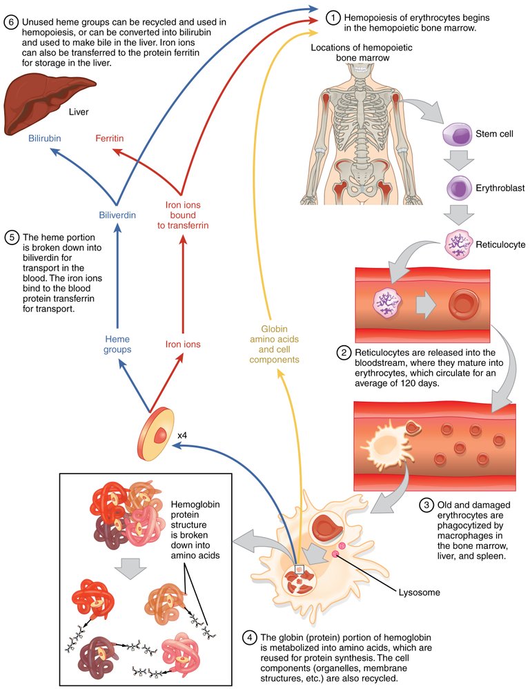
I explained in previous post the creation of Red Blood Cell, but I will further explain it in the life cycle of Red Blood Cell. Stem cells hemocytoblast in the Bone Marrow divides into Myeloid stem cells and lymphoid stem cells. The Myeloid Stem cells are responsible for erythrocyte. The Myeloid stem cells, produces proerythroblast, then to Basophilic erythroblasts, to polychromatic erythroblast, then Orthochromatic Erythroblast, then to Reticulocytes which doesn't have a nucleus, and then matures in circulation after 24 to 48 hours in the bloodstream into Erythrocyte. (You can read the formation in my previous post). From the snippet above, taken from my previous post, Erythrocytes have a lifespan of 100 and 120 days, which is a short lifespan, so understanding what happens and the complete process of the cell is important. Understanding the membrane of the red blood cell is important to knowing the process of Erythrocyte death.
The Erythrocyte Membrane Model consists of the spectrin tetramers (alpha and Beta), which is anchored by the Ankyrin at the actin junction, to the band-3 proteins, which attaches to the cell membrane. The Red Blood Cell, at its early age, is very flexible as a result of these cytoskeletal protein structure, thereby allowing it to reduce in size and formation, to be able to reach into smaller capillaries and places in the body, but as the Red blood cell ages, the protein starts to degrade causing the red blood cell to be rigid after which they get destroyed.
The destruction (Eryptosis), of the Red Blood Cell occurs in the reticuloendothelial system which includes the spleen, liver, and bone marrow, but the main site is the spleen where Macrophages are able to recognize and engulf them. In the Macrophage, hemoglobin is broken down into heme, and globin. The globin is degraded by proteases into amino acids, while the iron in the Heme is extracted. The Amino acid which is degraded, can be recycled for RBC production. The Iron from the heme binds with apoferritin to form ferritin. The ferretin forms together to become hemosiderin in the bone marrow (you can read that in my previous post).. In the same process, Heme is also oxidized to become biliverdin, and then converted to bilirubin which is bonded to albumin in the plasma and sent into the liver for bile creation and also sent to the intestine which would be excreted..
To explain the process of excreting the bilirubin, let me talk about Urobilinogen. The liver needs bilirubin for bile creation. The bile is sent to the duodenum, and then the large intestine, where the bilirubin is broken down to Urobilinogen by Bacteria proteases. A percentage of the Urobilinogen is reabsorbed into the bloodstream while some goes to to the kidney to be excreted with Urine. Urobilinogen is the reason for the yellowish color in urine. Some other part of the Urobilinogen gets broken down to become stercobilinogen which is excreated with feces causing the brown color in fecal mater. When feces isn't brown, it could sometime be as a result of the inability to secrete bile, which could be as a result of a gallstone. . The inability to pass out bilirubin in excretion would mean the bilirubin would be absorbed back into the bloodstream and excessive bilirubin in the bloodstream could lead to jaundice, which would cause the yellowing of the scela in the eyes and the body (palm) .
Conclusion
It is wrong to believe that the red blood cells in your body do not die, or they live forever. They actually have a life expectancy (or maybe I could call it an expiration date). Sickle cells die in a shorter period of time, which is often around 10 to 20 days, compared to 120 days which is expected to be the life span. This could lead to shortage of Red Blood Cell, causing Anemia.
Images
Excellent, It is very important topic in our medical community, greetings 🙋🙋🙋🙋
Thanks a lot for reading, I am glad you did
Great! Its amazing how our body works!
!1UP
Click this banner to join "The Cartel" discord server to know more.
Posted using Splintertalk
Thanks a lot @kwskicky
You have received a 1UP from @kwskicky!
@stem-curator, @neoxag-curator
And they will bring !PIZZA 🍕.
Learn more about our delegation service to earn daily rewards. Join the Cartel on Discord.
Thanks for your contribution to the STEMsocial community. Feel free to join us on discord to get to know the rest of us!
Please consider delegating to the @stemsocial account (85% of the curation rewards are returned).
Thanks for including @stemsocial as a beneficiary, which gives you stronger support.
Indeed our body is amazing. This is just beautiful.
You are right. Our body is a work of art and act of science