Visual Disoder (Macular Degenerative Eye Disease)
Having a degenerative eye disease can be very worrisome, allowing people to value everything they can see in their lives seeing that they do not know how many more suns they will see before their eyes degenerates completely. There are so many degenerative eye diseases that affect humans ranging from Glaucoma, Retinitis pigmentosa, Diabetic retinopathy, and many more. Today I will be describing Macular Degeneration, also known as Age-Related Macular Degeneration (AMD/ARMD). So let's begin without any further ado.
A lot of people with Macular degenerative disease complain of blurry visions, inability to see things unless it is upclose towards the eye completely. With age realted macular degeneration, the peripheral vision is working fine but the central vision is affected and with the dry macular degeneration, it would occur over the years as the person grow older if the person has the risk factor of having the disease. People with macular degeneration have Scetomas(blind spots), and they usually have night blindness (poor night vision), and blury color perception. The blindspot is not a completely blackout spot, the brain helps to fill in the areas that are not showing not as black but just an empty region. Macular degeneration accounts for about 8.7% of all types of worldwide blindness.
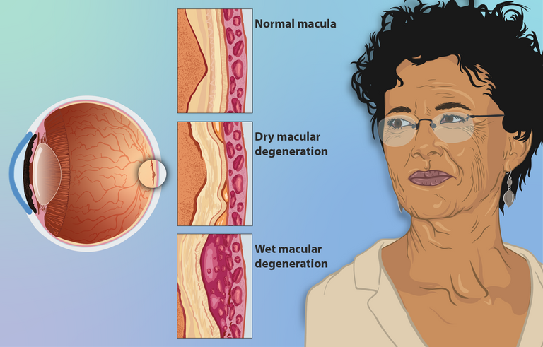
Age-Related Macular Degeneration is a degenerative disease just as its name implies. It is a degenerative disease that is associated with Central vision loss of the eye. The eyeball is made of the cornea which is the outside portion of the eyeball, followed by the iris which is the color pigment of the eye. If a light ray passes through the pupil, it reaches the lens where the light hits the retina which sends the information to the optic nerve which sends the information to the brain. At the back of the eye are the Choroid and the Sclera. Within the retina is the Macular which is the bulls eye of vision that contains cones. In simple terms, Macular degeneration has to do with the macular having issues, leading to central vision problems.
The macular can be affected in two ways which are the wet (atrophic) and dry (Exudative) ways. In the dry (atrophic) macular degeneration, there is an accumulation of the Drusen proteins, separating the choroid from the retina leading to retina atrophy and leading to degeneration. Dry macular degeneration is the most common type of macular degeneration as about 9 out of 10 people with macular degeneration suffer from dry degeneration. It is the decrease in the profusion to the retina which is a slower change to the vision. It has to do with the ischemia of the macular, causing a blockage of the microvasculature. This process leads to a separation of the brunch membrane and the retina pigment epithelium (RPE).
In the wet type of macular degeneration, the choroid starts to produce new blood vessels which are thin and weak, and have the potential to leak. When they leak, they release blood, and fluid which then separates the macular forming a hemmorhagic Edema scaring. The wet macular degeneration is exudative and less common compared to the wet. This degeneration occurs fast and it is often noticed at any age. The abnormal growth of the new vessels are thin and weak leading to a perfuration of the vessel allowing blood and fluids to accumulate beneath the macular leading to a scar which would affect the transmission of visual information to the brain leading to macular degeneration.
RIsk factors of Macular degeneration can be divided into modifiable and non-modifiable. The non-modifiable one is Age and Family history, as these cannot be modifiable. Other risk factors which are modifiable include smoking, Poor intake of vitamins that can help with visual quality, and hypertention. This can be modified by smoking ceasation, use of supplements, and laser therapy and injections to help treat the degeneration. Symptoms are usually blurry visions and metamorphopsia (distorted vision). Using Fluorescien dye to access the blood vessels and Fundoscopy to view book of eye are the ways to diagnose. Interventions to patients with macular degeneration would include modifications which includes givinng glass, improved lighten, occular injection and laser therapy.
One of the causes most of the time can also be traced to genetics
Sure, but it usually comes with old age in some case.
Congratulations @elity-sitio! You have completed the following achievement on the Hive blockchain And have been rewarded with New badge(s)
Your next target is to reach 100 posts.
You can view your badges on your board and compare yourself to others in the Ranking
If you no longer want to receive notifications, reply to this comment with the word
STOPTo support your work, I also upvoted your post!
Check out our last posts:
The most serious cases that I don't want to hear is if a disease is related to Genetics. Its healing is very complicated and in most cases, they are impossible as you rightly said.
It looks like a lot of diseases have a form of genetics in them, making it hereditary in one way or the other. This goes from diabetes, high blood pressure and so on. Most conditions tend to be genetically transferable.
Yay! 🤗
Your content has been boosted with Ecency Points, by @elity-sitio.
Use Ecency daily to boost your growth on platform!
Support Ecency
Vote for new Proposal
Delegate HP and earn more
I used to think that AMD comes with all age and that everyone who gets to a certain age is at risk but now, I feel it's more hereditary
It is actually both depending on the condition.
Okey dokey
Thanks for your contribution to the STEMsocial community. Feel free to join us on discord to get to know the rest of us!
Please consider delegating to the @stemsocial account (85% of the curation rewards are returned).
Thanks for including @stemsocial as a beneficiary, which gives you stronger support.