How to differentiate an animal blood from a human blood: its importance in forensic science
So today, we are going to discuss something you probably never saw coming your way.
This will be an impactful read. I could remember vividly during my time in the university, I always had a lot of thoughts when I read textbooks, one of which I at some point had to stand up during lectures to inquire from the lecturer. I could remember asking one of the histopathology lecturer this - "how do we know that a blood is from a animal or a human being".
The question looked surprising to her, because from her reaction, I could guess she wasn't expecting the question. It was virtually off course to what we were learning at that moment, though something spurred me into asking her that question then, can't exactly remember right now. Pardon my curiosity, I couldn't just hold it. Did I get an answer to my question, an emphatic NO, I didn't. Reason till date I can't actually explain.
Anyways, don't worry about my case, I now have a better understanding through professional experience and from personal research. Today after reading this article, you sure will be able to confidently distinguish animal and human blood samples.
Is my question just a random question or does it have no useful importance in any way. Reading this article will give you a broader understanding. We will begin the article proper by taking some useful attributes that will help you easily identify the blood as either, of animal origin or human. Sit tight and enjoy.
Respiratory pigment
Most blood of animals have one thing in common and that is, a respiratory pigment. Human blood contains 4 polypeptide chains of haemoglobin as respiratory pigment and this binds 4 Oxygen molecules. Haemoglobin is bound to iron in human. It is mainly responsible for the transportation of oxygenated red blood cells to various parts and organs of the body system.
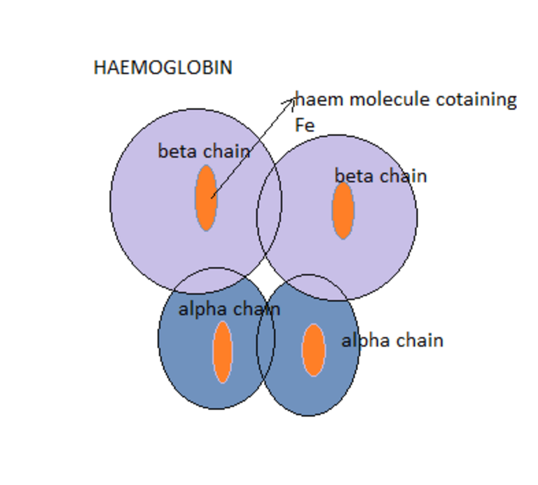
Haemoglobin pigment type found in human blood
Without the binding of haemoglobin to oxygen, it will difficult for end organs to receive oxygenated blood from the lungs. So in essence, respiratory pigment is the engine of the vehicle (blood) for effective transportation of nutrients and substances around the animal or human body.
In animals, the case is different because they have not just one but 4 respiratory pigments namely - Haemoglobin (present in mammals), Haemerythrin (found in marine invertebrate animals like the annelid worm, Haemocyanin (they contain copper and found in arthropods and molluscs) and Chlorocruorin (This is violet in colour and mostly found in Marine polychaete worms.
So on examination of animal blood, you wouldn't be expecting to see the usual specific red coloured haemoglobin you see in humans, animals have, but theirs is different. It is worth knowing at this point that, it is the haemoglobin that confers the colour of blood.
In humans, the blood is mostly red because type of haemoglobin found in us while in animals, it varies. In essence, this can also be used as a distinguishing factor.
Next we want to discuss is the shape of the red blood cells, a very important distinguishing feature.
Shape of the blood cells
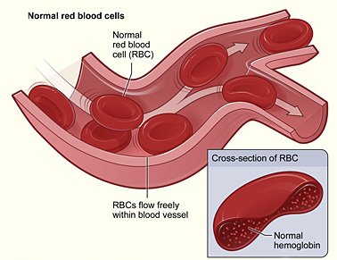
The red blood cells are the major component of the human blood. In essence, you see them more in abundance under the microscope. In humans, the shape of the red blood cells are biconcave and round with central pallor.
Presence and absence of nucleus
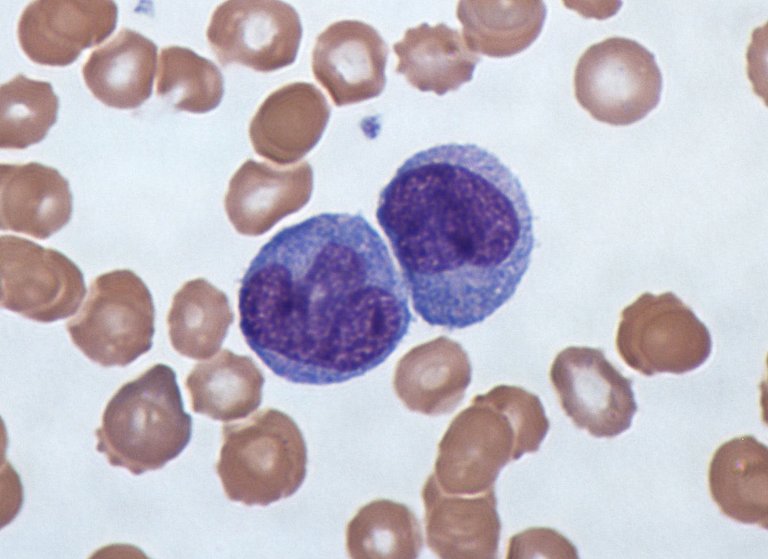
Very importantly, matured human red blood cells are usually enucleated (they don not have nucleus). Note this, the presence of nucleus in any matured red blood cells of humans is a serious disease condition and one of which is likely cancer (leukemia).
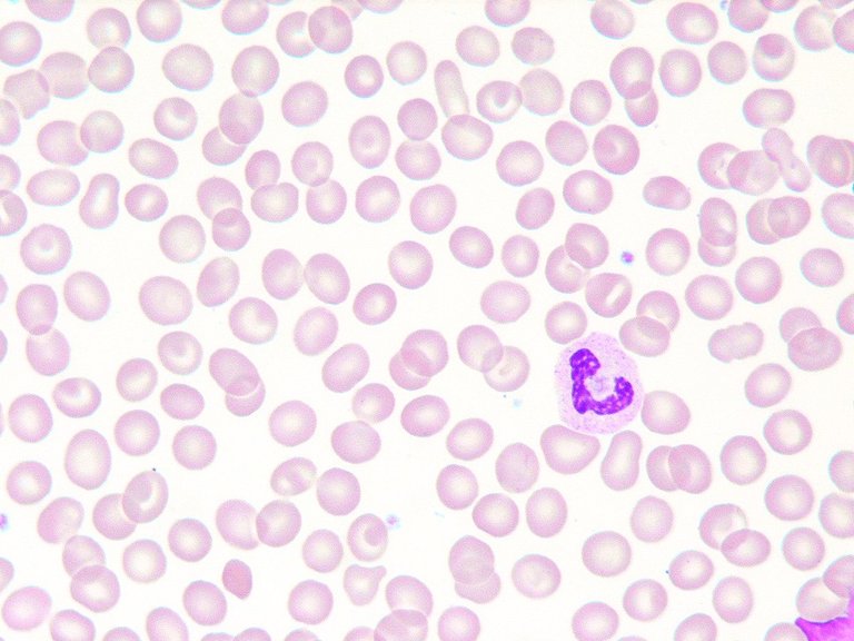
Matured human red blood cells with its visible central pallor
The image above shows the numerous red blood cells with one white blood cell (Monocyte) located around 3'clock. Nucleus are only found in reticulocytes (immature red blood cells). So any point you find nucleus in human blood and you have ruled out the fact that it is not a reticulocyte, the only thing that should come to your mind is a disease condition.
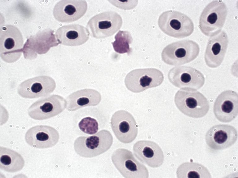
On the other hand, blood from animals (mostly higher ones) are always nucleated (they have nucleus).
Make no mistake about it, the nucleus is a very important structure found in cells because it houses the genetic machinery of cells. One question you may want to ask, since nucleus is that important, why does the human matured blood lack it?
Mature red blood cells (RBCs) do not possess nucleus along with other cell organelles such as mitochondria, Golgi apparatus and endoplasmic reticulum in order to accommodate greater amount of haemoglobin in the cells. However, immature red blood cells contain nucleus.
Going by one of the functions of human red blood cells, they transport mostly oxygen to various parts of the body as well as to vital organs and tissues. Oxygen is strongly bond to haemoglobin in the red cells and this haemoblobin occupies almost the surface area of the red cells. The nucleus in the red cells will disrupt its full functionality at maturity.
The red blood cells wont be able to effectively carry out its role of binding to oxygen and delivering it to various parts of the body. Moreover, in addition to the above reason, having nucleus will prevent the red blood cells from maintaining its bi-concave shape, a very important feature that helps it movement within the blood vessels (the vein, artery, capillaries, venules etc)
The sizes also matters
Another unique distinguishing factor between animal and human blood is the varying shapes. The matured red blood cells in humans measures approximately 7-8 micrometer and a thickness of 2-3 micrometer. If you noticed a trend in the image below, all the animal blood are bigger in diameter as compared to the human blood.
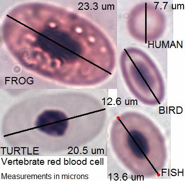
Human and animals (vertebrae) red blood cells diameter
It is not surprising because, the red blood cells have very high special functions. They will need to maneuver alot through the blood vessels and especially through the spleen which has a very narrow entry point.
Just like we earlier explained that, animal red blood cells have nucleus. By mere looking at all the images in the general image, you would observe that they all have a darker centrally located structure. This is referred to as the nucleus while the human matured red blood cells had no nucleus.
Matured red blood cells are able to pass into the spleen by their explicable high ability to bend, twist and reform their shape while passing through. A large red blood in human will only end up clogging blood vessels.
Twisting and reforming ability of red blood cells as it passes through capillaries
Have you ever wondered what happens to red blood cells after reaching about 120days? Perhaps you know what happens to them, do you know how it happens and what initiates the process? Well you are about to know now.
Red blood cells have a half life of only 120days and after which they and recycled. As red cells move round the blood vessels, they age while acquiring various impurities that are attached to their cell membrane. The more this impurities are attached, the more they loose their twisting ability and also slightly increase in size.
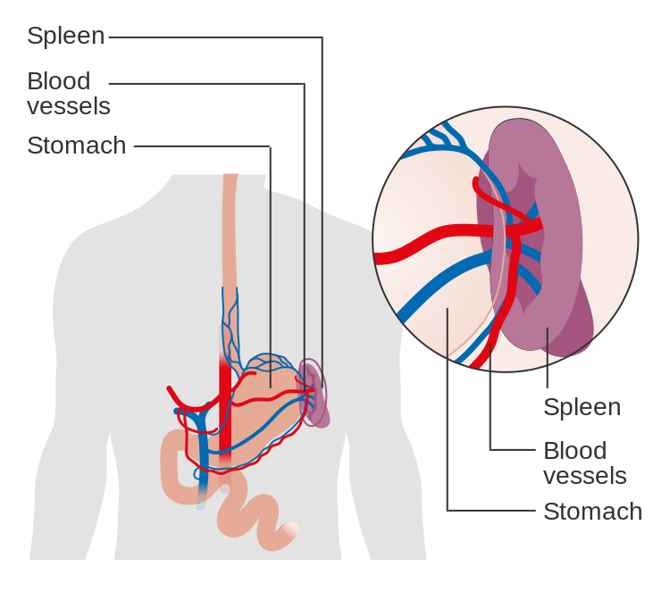
The spleen is the police check point for all red blood cells that circulate round the human blood vessels. It the major organ that determines the size, shape and as well as the health status of any red blood cell.
It primarily plays the role of clearing red blood cells that have variation in size, shape and those that have lost their ability to deform or twist.
Red blood cells with such features are seen as unhealthy and are thus cleared. I.e destroyed, and ultimately it's content like the Protein - globin and and iron content - haem recycled for further formation of new red blood cells.
The simple mechanism of the spleen destruction of unhealthy red blood cells lies in its aperture, through which the red blood cells passes.
Passage route of red blood cell through the spleen
Normal size shaped healthy red cells can twist, deform and safely pass through this narrow route but unhealthy res cells cannot, hence, they are trapped inside the spleen for destruction. This is the simple summary of how aged red blood cells are recycled.
The cellular component
Examination of the human blood under the microscope will reveal the other types of cells besides the red blood cells. E.g white blood cells (which includes the eosinophils, neutrophils, monocytes, lymphocytes and basophils), platelets etc but in animals, the case could be sometimes different. For emphasis sake, their only 5 types of white blood cells in human blood but animals have varying types.
The cellular component in animals besides the varying white blood cells is the haemocytes (cells in animals that play protective role - immunity, just like in human where the white blood cells fight against foreign pathogens invading the body system).
Clinical Biochemical tests
A simple test that can also help you know the type of blood you are in possession of is to simply run a glucose test. Research has shown that glucose distribution pattern in humans and animals is quite different. For example, in humans, most of the glucose content of the blood can be seen almost evenly shared between plasma and the red blood cells. The red blood cells contains about 42% of the entire circulating glucose in humans while the remaining 58% is found in the plasma.
On the other hand, in animals, the case is different because almost all the glucose content is located within the plasma while the remainder is found in the red blood cell. It is not surprising because humans as higher animals, the red blood cells are much more advanced in structure and functionality and as such will require enough energy source in the form of glucose.
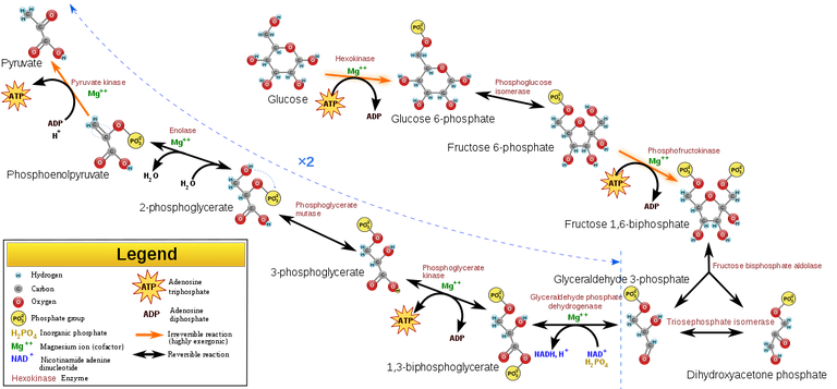
Glycolysis - pathway through which sugar in the form of glucose is broken down
Ever wondered why Sodium fluoride-potassium oxalate, aka fluoride-oxalate bottle (yellow cap bottle) are used for blood glucose measurement? Is simply because fluoride oxalate inhibits enolase, the penultimate enzyme required in glycolysis (glucose degradation) for the conversion of glucose to pyruvate.
Note, red blood cells use glucose as an fuel for their metabolic activities. Through the process of glycolysis, they breakdown glucose and in the process, adenosine triphosphate (ATP) is released. The ATP is the major usable form of energy by the cells.
If this reaction is not stopped, the red blood cells inside the blood sample already collected will use up the whole glucose until their virtually nothing to measure. In this case, if you do the biochemical investigation, the result would either read extreme low blood sugar or zero. This obviously would lead to wrong diagnosis.
Is it possible to differentiate between animal blood from human blood physically
It is a near impossibility to differentiate between animal blood (higher ones) from human blood physically. Blood cells are microscopic in nature, meaning you cannot see them with an unaided eyes. The only way they are seeing is with the use of a microscope.
So in essence, it is quite impossible to fully differentiate the two without using microscope. If you are presented with two blood samples for differentiation and identification, the procedure below will guide you adequately if you are indisposed to advanced equipment to help you out.
How do you examine the blood cells microscopically?
The first thing you need to do, is to place a drop of each blood sample of blood on different grease free labelled slides. Make a thin blood film of each blood sample and air dry them. You then stain the blood film using any Romanowsky stains (Giemsa, Jenner, Wright, Field, May–Grünwald and Leishman stains). This stains contains two major chemical components (methylene blue and eosin) that typically differentiates the components of the cell into two namely - the nucleus and the cytoplasm.
The eosin is by nature an acidic dye and as such will bind and stain only the basic (alkaline) cell component like the cytoplasm on the other hand, the methylene blue being a basic dye will stain any acidic component of the cell. e.g., the nucleus.
To visualize the cells, you then use a binocular compound light microscope. Since the slide is stained, you need to allow sufficient light to pass through the glass slide and to do this, you totally open the condenser aperture and at the same also increase the light intensity.
Next thing you do is to set the nose piece of the microscope to X100 (Oil immersion lens). Place a drop of immersion oil on the stained film and place it on the slider of the microscope and the bring into focus the oil immersion objective lens by making sure it comes in contact with the immersion oil you already dropped on the film. You do this using the coarse adjustment knob. Finally, you then use your fine adjustment knob to fine tune the image for clearer visibility.
Under the microscope, you would be able to easily see some of the features we have elucidated on above, with the except of haemoglobin as it requires a more complex technique and equipment to be able to visualize it. But with the microscope alone, you should be able to easily know whether a blood sample is from an animal or from a human.
Blood film with Giemsa stain. White blood cells (center) surrounded by red blood cells
.jpg)
Remember, the easily identifiable features of a human blood is the presence of its unique blood types (red blood cells, white blood cells, and platelets) and then their shapes. From the image above and to be specific, the particular type of white blood cell seen is the monocyte because of its unique characteristic of having nucleus that almost completely covers the entire cytoplasm of the cell. All cells have their unique identifier.
Bloodstains found at crime scenes are very important evidence and one of the main sources of DNA. The two most common confirmatory assays for blood are the Teichmann and Takayama tests, which are both microcrystal tests.
In conclusion, the ability to differentiate between animal and human blood is very important especially to forensic experts. Knowledge of this will help in solving crime cases, accident cases involving hit and run drivers and more. It is wroth knowing at this juncture, all the above described techniques and methodology form the preliminary investigations that can be done to differentiate human blood from that of animals.
There are more advanced and sophisticated machines that differentiate human blood from that of animal and they mainly take advantage of the haemoglobin content of the blood. Remember we earlier established that, there exist some significant difference that exist among the various haemoglobin found in animal and human blood. These differences can be identified mass spectroscopy and even high performance liquid chromatography.
Since we may not be able to easily access these very expensive machines that require high level of expertise, we simply make do with our microscope, the microscopic eyes of diagnosis.
Till I come your way again, stay awesome.
References
Blood from humans and animals is different
Identification of species specific hemoglobin by isoelectric focusing
Discrimination between human and animal blood by attenuated total reflection Fourier transform-infrared spectroscopy
New Forensic Technique Quickly Tells Human Blood from Animal Blood
What a read! I never knew there were so many ways to tell how human blood differs from those of animals.. Thanks for sharing such a detailed post @cyprianj!
They both differ in so many ways... going deeper with advanced machines reveal alot.
Technology remains vital in propelling us to understanding life.. You have access to all these machines in your institution?
As the need arise, the machines are made available.
Definitely an interesting and informative read. I will go through it again.
Are there no exceptions whatsoever? Like nothing else in the animal kingdom is even close to having those same properties of human blood cells?
Definitely none, they may have close resemblance but never exact. That makes us whole lot advanced.
What about vampires?
Haha, do those really exist. I still believe vampires are myths.
Well I guess their blood would be easy to test: just put it under sunlight and see if it catches on fire!
Haha, that's definitely the best experiment.
Great Post!
!1UP
Thanks for reading
You have received a 1UP from @luizeba!
@stem-curator, @vyb-curator, @pob-curator, @neoxag-curator, @pal-curatorAnd they will bring !PIZZA 🍕
Learn more about our delegation service to earn daily rewards. Join the family on Discord.
One reason why I would never make a good doctor is because I can never stand the sight of blood, but this is a lot of eye-opening information, thank you, @cyprianj.
Lol...not in all cases. Sometimes, you get used to seeing them.
Thanks for your contribution to the STEMsocial community. Feel free to join us on discord to get to know the rest of us!
Please consider delegating to the @stemsocial account (85% of the curation rewards are returned).
Thanks for including @stemsocial as a beneficiary, which gives you stronger support.
Your content has been voted as a part of Encouragement program. Keep up the good work!
Use Ecency daily to boost your growth on platform!
Support Ecency
Vote for new Proposal
Delegate HP and earn more
So, blood is not just blood… This is a good thing to learn, which I didn’t know before reading this blog.
Just one question, although I think I guess the answer correctly (but better to ask the expert to be sure ;) ). In fact, just two related questions...
You mentioned that animals have 4 respiratory pigments in their blood. Do they all have 4 pigments, or do they have up to four pigments? Similarly, do animals always have a given type of white blood cells, or could their blood feature admixtures of several types of white blood cells? Thanks in advance for answering me!
Cheers and have a good week!
They have different pigments. All the four pigments cannot be in one animal. It is the varying pigments that gives their blood different shades of colour - green, yellow, violet etc on the other hand we humans have majorly red colour conferred by haemoglobin.
Regarding the white blood cells, definitely theirs vary from that is humans though not Much. In animals, theirs is mostly called haemocytes (also function like wbc in Humans) case in point animals that have a closed circulatory system (blood system made up of a heart pumping Blood through blood vessels) like Humans but in none mammals.
In summary, animals have similar wbc as humans depending on specie and secondly the difference can easily be detected through their quantity. Wbc in animal are more abundant than in humans, just the way glucose is found more in the plasma of an animal than in humans.
Thanks for those clarifications. They are perfect and allow me to adjust how I thought about the situation.
Cheers!
Hello @cyprianj This is alfaz and i want know how to post on this stemgeeks platform. I'm knowladge of medical field and i want post related to this but would you please tell me how to do it any tutorial for this?
@stemsocial
You mean Stemsocial, all you need do is to select Stemsocial as the community you would love to post in and upload. that's all.
Ok so I should have just open hive.blog and open stemsocial community and then create a post so that post uploaded so stem social community and website.
Yeah, that's just it. Select the Post to be posted on Stemsocial community.