Cerebral angiography⛑️⛑️
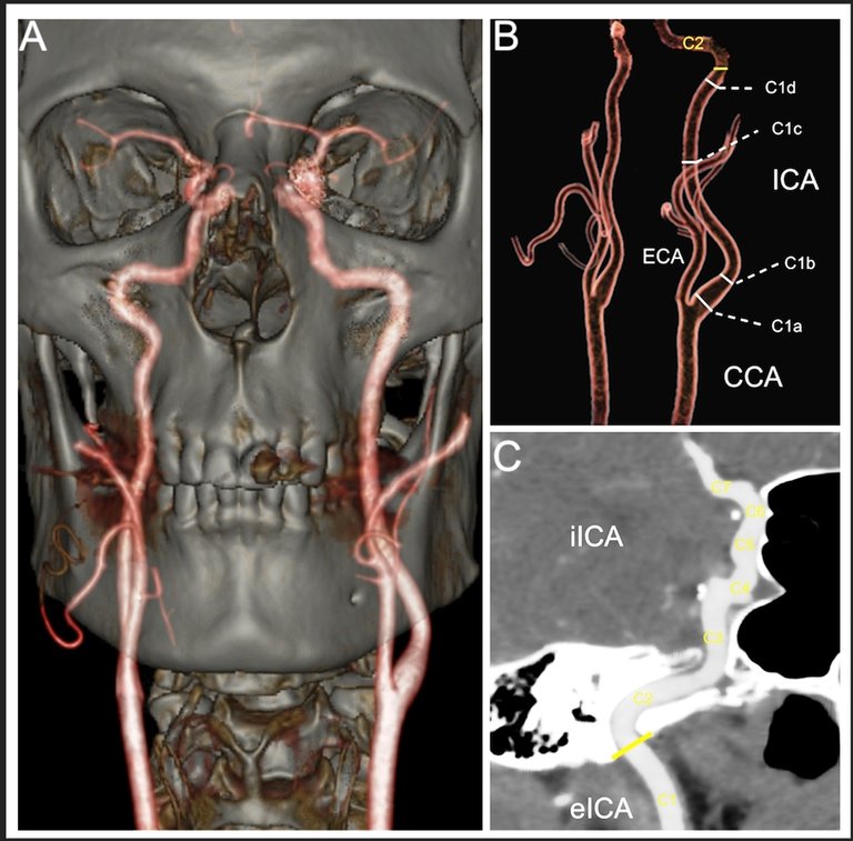
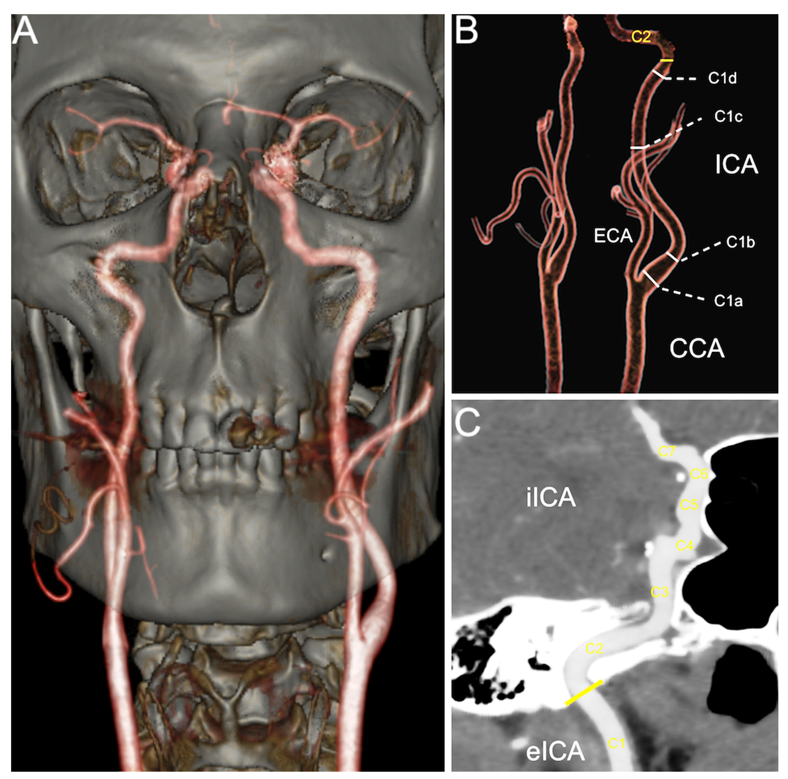
Hello 🙋🙋🙋🙋friends of the #StemGeeks community today I want to talk to you about one of the most important diagnostic means in our hospital center, cerebral angiography (Fig. 1), frequently used in patients with brain disorders, for diagnostic and sometimes therapeutic purposes.
Source: MDPI
This is a medical evaluation technique that allows the study and analysis of the blood flow of the cerebrovascular system. This is a technique in which X-rays are used to visualize the flow and state of the circulatory system by injecting a contrast into the main cerebral blood vessels. The images obtained are generally very clear and make it possible to accurately identify disturbances in the blood circulation of the brain.
Procedure
⛑️⛑️⛑️⛑️⛑️⛑️The patient is placed on the X-ray table, his head is immobilized and a sedative is administered, at the same time his cardiac activity is monitored, then the patient is inserted a catheter (Fig. 2) into the arteries of the arm or leg, which will be guided by the artery to the neck with the help of X-rays. Once there, a contrast solution is injected through the catheter to later take images of the blood circulation using X-rays.

Source: wikipedia
After the X-rays are taken, the catheter is removed. Pressure is immediately applied to the leg at the insertion site for 10 to 15 minutes to stop the bleeding or a device is used to close the small hole. Subsequently, a tight bandage is put on. The leg should be kept extended for 2 to 6 hours after the procedure. The area should be observed for bleeding for at least the next 12 hours.
It is important to note that during the examination you may have brief pains and pressures as the catheter moves through the body, in addition to burning or hot sensations in the head when the contrast dye is administered, but this disappears after a few seconds.
Types of cerebral angiography
⛑️⛑️⛑️⛑️⛑️⛑️Cerebral angiography is a technique that has several variants depending on the mechanisms used to assess the condition of the patient's blood vessels. Some of the best known are the following.
- Conventional angiography (by intraarterial digital subtraction).
This is the previously explained procedure, in which the catheter is placed in the artery and guided to its target. It is an invasive procedure that is usually the most common due to its effectiveness and the high level of sharpness it allows. The catheter is usually inserted femorally (Fig. 3) through the groin to the aortic arch, where after a first injection of contrast, the catheter is placed in the artery to be analyzed.

Source: researchgate
- Helical computed tomography angiography (TC).
In this case, no type of catheter is introduced into the subject's body, but it does require the injection of a contrast in order to obtain the image by CT. This one is less invasive than the previous one.
- Magnetic resonance angiography.
In this type of angiography, no catheter is used either, not being an invasive technique. It involves performing an MRI, not using radiation as in other cases.
Uses of this technique
⛑️⛑️⛑️⛑️⛑️⛑️Cerebral angiography is a test that is still used today as one of the main diagnostic methods for observing the circulatory flow and the state of the blood vessels of the brain. There are multiple disorders and diseases that the application of this technique allows to observe.
- Cerebrovascular accidents or strokes (Fig.4)

Source: mayo clinic
Angiography allows to observe the existence of extravasation and ruptures of blood vessels, or the absence or obstruction of circulation in some area of the brain. That is why we are facing a valid technique both to detect ischemia and to visualize cerebral hemorrhages.
The use of angiography makes it possible to detect the presence of aneurysms, bulges of the arterial wall filled with blood and relatively weaker wall that can rupture.
- Tumor
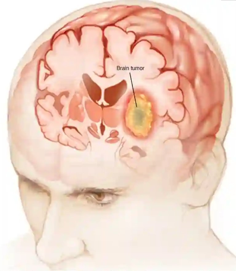
Source: mayo clinic
The presence of tumors in the brain tends to cause disturbances in the blood flow of the brain, as well as provoke phenomena such as strokes. Therefore, angiography allows to observe the presence of abnormalities generated by the presence of tumors.
- Malformations
The existence of congenital malformations, can also be detected by this technique.
- Arterial or venous alterations
Using cerebral angiography, it can be observed if the blood vessels of the brain are in a good state of health, if they are inflamed or if disorders such as atherosclerosis occur.
It is also used to assess whether or not there is brain death. Specifically, it is evaluated whether or not there is blood flow, observing an absence of irrigation in those cases of brain death.
- Other disorders
There is the possibility of observing through cerebral angiography the presence of different disorders and diseases apart from those previously mentioned. For example, vascular alterations may be found in the presence of neurosyphilis.
How to prepare for the exam
How to prepare for the exam
⛑️⛑️⛑️⛑️⛑️⛑️Always before the procedure you will be examined by the attending physician who will order the performance of complementary examinations to assess your general condition.
It is important that you inform the doctor during the consultation if you suffer from any disease, if you have a history of bleeding, if you are allergic to the X-ray dye that will be used, if you are allergic to Iodine, if you suffer from any kidney disease since this way is that the radiological contrast that will be used is eliminated, if you are pregnant since most of these diagnostic tests can cause damage to the fetus with the future appearance of congenital malformations.
You must sign a consent form, which explains the procedure and the possible complications that may appear during its completion. When you arrive at the test site, you will be given a hospital gown to wear and must remove all jewelry.
Benefit
⛑️⛑️⛑️⛑️⛑️⛑️Angiography can eliminate the need for surgery. If surgery is still necessary, it can be performed more precisely, it allows the taking of very detailed, clear and accurate photograph of the blood vessels of the brain, these are more accurate than those produced by carotid Doppler ultrasound or other non-invasive imaging methods of blood vessels, in addition the use of a catheter makes it possible to combine diagnosis and treatment in a single procedure.
Risks and possible side effects of this technique
⛑️⛑️⛑️⛑️⛑️⛑️Cerebral angiography is a generally safe technique that does not tend to cause complications, but this does not prevent it from having risks and adverse side effects that can cause alterations of varying severity.
One of the risks is the possibility that the patient has an allergy to the applied contrast (usually iodinated). Also, this could cause discomfort or even destruction of some tissues if it is extravasated outside the vein. It can also be risky or harmful for people who suffer from kidney problems or diabetes.
The existence of symptoms such as tingling, breathing difficulties, vision problems, infection of the route through which the catheter has entered, control problems of the limb in which it has been inserted, speech problems or hemiparesis are a sign that there may be some type of complication to be treated quickly.
Finally, special caution is necessary in the case of pregnant or lactating women, since the emitted radiation could be harmful. It can also happen that a tear of the artery is caused that generates some type of hemorrhage or clots that can plug the vessel, although it is something very uncommon.
After the procedure
⛑️⛑️⛑️⛑️⛑️⛑️Depending on which site was used to inject the contrast dye, you need to lie in a recovery room for several hours after the procedure. If the groin, arm or leg was used, on the side of the injection site it will remain straight for up to 12 hours. If an area of the neck was used, it will be monitored for signs of hoarseness, shortness of breath, or pain difficulty swallowing.
A nurse will monitor your vital signs, neurological signs, and injection site. While you are in the recovery room, you may be given medication for pain or discomfort related to the injection site.
You will be advised to drink water and other liquids to help remove the contrast dye from your body. You can resume your normal diet and activities after the procedure unless your doctor decides otherwise.
When you have completed the recovery period, you may return to your hospital room or be discharged home. If this procedure was performed on an outpatient basis, you will need to provide for someone else to take you home.
Instructions for home
⛑️⛑️⛑️⛑️⛑️⛑️Once at home, the injection site should be monitored for bleeding. A small bruise is normal, drink plenty of fluids to avoid dehydration and to help remove the contrast dye, you should not do strenuous activities or take a hot bath or shower for a period of time after the procedure.
It is important that you tell your doctor if you have any of the following symptoms:
Fever, chills, increased pain, redness, swelling, bleeding or other discharge from the injection site in the groin, feeling cold, numbness, tingling, or other changes in the affected limb
Finding
Summarize
⛑️⛑️⛑️⛑️⛑️⛑️All diagnostic and therapeutic means are very important, thanks to them we can make the diagnosis of diseases that are in the insides of the organism, which through physical examination by the doctor would be impossible to diagnose.
At the same time, to say that all the tests are not 100 percent safe and all have the risk of producing unwanted effects on the patients to whom they are applied, serious accidents can even occur that ruin the patient's life.
The use of angiography is necessary and thanks to it it has been possible to increase the life expectancy of patients suffering from any disease in which its use is indicated.
Well friends I hope you liked the post, that it is useful to you, thanks for reading and dedicating some time, have a great week and until next time.
Bibliographic references
- Camargo, M.; Peralta, A.; Arias, W.; Mercado, C.; Laguna, Y.; Cuellar, J.; Laforcada, C.; Paz, G.; Durán, J.C.; Aramayo, M.; Fortun, F. & Núñez, H. (s.f.). Stroke Protocol. Bolivian Society of Neurology.
- https://hospitalgalenia.com/que-es-la-angiografia-cerebral/
- https://psicologiaymente.com/clinica/angiografia-cerebral
- Daroff, R.B.; Jankovic, J.; Mazziotta, J.C. and Pomeroy S.L. (2016). Bradley's Neurology in Clinical Practice. 7th ed. Philadelphia, PA: Elsevier.
- https://medlineplus.gov/spanish/ency/article/003799.htm
- https://www.radiologyinfo.org/es/info/angiocerebral
- https://hospitalgalenia.com/que-es-la-angiografia-cerebral/
- https://www.topdoctors.com.co/diccionario-medico/angiografia-cerebral
- Millán, J.M. & Campollo, J. (2000). Diagnostic value of cerebral angiography in the confirmation of brain death. Advantages and disadvantages. Med. Intensive, 24(3); 135-141. Madrid.
⛑️⛑️⛑️⛑️⛑️
⛑️⛑️⛑️⛑️
⛑️⛑️⛑️
⛑️⛑️
⛑️
Congratulations @lindoro! You have completed the following achievement on the Hive blockchain and have been rewarded with new badge(s):
Your next target is to reach 3000 upvotes.
Your next target is to reach 400 upvotes.
You can view your badges on your board and compare yourself to others in the Ranking
If you no longer want to receive notifications, reply to this comment with the word
STOPTo support your work, I also upvoted your post!
Support the HiveBuzz project. Vote for our proposal!