What's in the Slide? Guess and Win #4 [CLOSED]
Guess what's in the slide? It's exactly what it says. Question is at the first part of the post. Instructions at the second part of the post.
Winner: @nikv
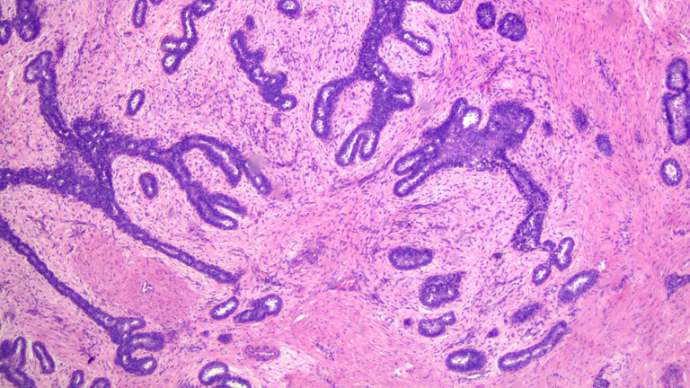
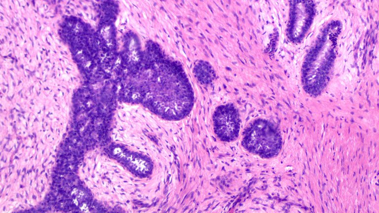
Answer: Fibroadenoma
Bonus Answer: Phyllodes Tumor
Bounty 2 HBD
It's a common neoplasm on women during child bearing age. One differential includes the Phyllodes Tumor which can be quite tricky to clinch sometimes as they present with almost the same morphologic findings.
You would see the epithelial component compressed and these would look like leaf patterns. There are two main proliferations here, the intercanalicular and intracanalicular where some tumors can have a predominance of one pattern. The pink areas are the stromal areas with fibrosis (showing intercanalicular proliferation) think of this pattern as constricting the lobules as they grow.
Taken at Scanner view (40x magnification).
Taken at Low power view (100x magnification).
Specimen: Breast from a woman within the child bearing age range.
Question: What's the possible diagnosis? There are two possible answers to this.
Bounty: 1 HBD
Bonus: Another 1 HBD to whoever guesses the second possible answer. You can win twice.
The first one that guesses this right gets that HBD. The prize gradually increases when no one gets it right for each successive post on the series. At the very least, each post has a 1 HBD bounty on it. I'll prioritize adding more bounties on older questions compared to new questions in the series.
The mechanics of the guessing game:
I post the images and give a small detail about it with a corresponding question.
Comment the answer and whoever gets it right first wins the prize. There's a time stamp on the comment section so it's easy to determine the winner if multiple users got it right.
In the event that no one gets it right, the contest will still be open indefinitely. Feel free to ping me if you backread the previous posts in the series.
I'll add new conditions to the game as needed.
You are free to Google for answers or use whatever means you got at your disposal with a corresponding reason why you think it is so. It's easy to get it right by throwing words around so I want to see whether you studied the image.
Make as many attempts as you want. The only time an attempt isn't allowed is when the contest has been closed. There can only be one winner per post. You can try multiple times but spamming some answers from a bucket list isn't going to be get you a reward.
Since you have the advantage of googling the answer. I'd be requiring a short explanation why. It doesn't need to be the exact rationale but if you're close enough I wouldn't mind. This is to prevent anyone that just answers and win through dumb luck, I'd like to see some conviction on the answers.
Note:
I'll be copypasting the mechanics of the game including this line so that anyone who is new to the series wouldn't have to click more links just to backtrack what's going on. If anyone wants to cry I'm milking the reward pool by posting copy paste posts, understand the images here are from actual cases where I took the time to have them recorded.
I highly encourage you to research the answer in the hopes you can actually learn from the experience. Pathology is fun.
I'm also confident you can't find any image that match exactly as the ones I'm sharing because these are from my personal study slides. You can of course see similar images because they can show the same histomorphologic findings you'd expect from the specimen.
Good Luck!
Case 1 Closed
Case 2 Closed
Case 3 Closed
If you made it this far reading, thank you for your time.
Posted with STEMGeeks
Ameloblastic Fibroma
You're off with one word. See nikv's answer.
Benign Fibroadenoma
or epithelial hyperplasia and stromal hypercellularity
Nice, there's still the bonus question of other possible diagnosis for this. Will wait for that answer before closing the thread.
Second possibility: sclerosing adenosis
I could force that answer as a third possibility as a differential but the other answer in mind is a closer bet in morphology. Sclerosing adenosis would have calcifications and apocrine metaplasia missing. It's a close candidate.
Phyllodes tumor is the differential and damn I forgot to add histology to my search and now I can't unsee those images on google
Best guess - Papillary carcinoma
No, there's nothing in the slide that has a papillary architecture.
Fibroadenoma
Correct, but it's already answered by @nikv.
There's still another possible answer I'm waiting.
Congratulations @adamada.stem! You have completed the following achievement on the Hive blockchain and have been rewarded with new badge(s):
Your next target is to reach 50 comments.
You can view your badges on your board and compare yourself to others in the Ranking
If you no longer want to receive notifications, reply to this comment with the word
STOPCheck out the last post from @hivebuzz:
Support the HiveBuzz project. Vote for our proposal!