What's in the Slide? Guess and Win #2 [CLOSED]
Guess what's in the slide? It's exactly what it says. Question is at the first part of the post. Instructions at the second part of the post.
Winner: @nikv with "Biliary obstruction caused by parasites with schistosoma in an unusual location"
Answer: Schistosomiasis spp. infection
2 HBD Bounty for this case.
Let's go over the case:
This was signed out as findings consistent with Schistosomiasis spp. infection. We didn't declare the specific species because the method of collection isn't the optimal way to demonstrate the parasitic ova.
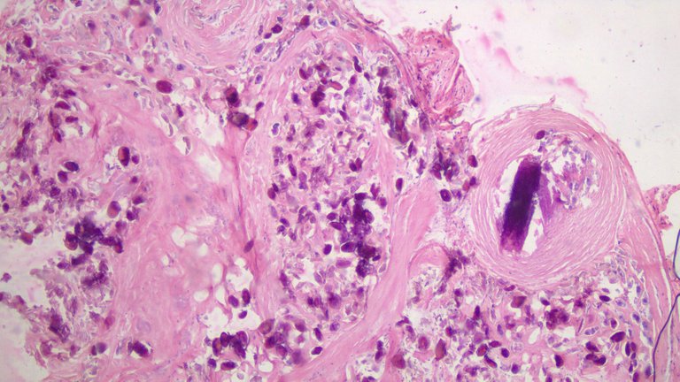
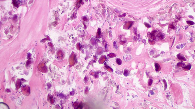
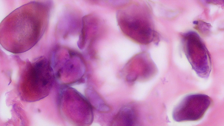
The "omental nodule" was a fibrotic and calcified tissue where the surrounding tissue tried to contain and react to the chronic infection. The purple color here is how calcium stains under Hematoxylin and Eosin stain preparation. I tried to find a group of ova that can show some clear delineation for between the shell and what's inside it.
Leyte is an endemic area for Schistosomiasis cases. The patient travelled to our region and subsequently sought consult to our institution. We usually report these cases to the local health regional center for monitoring. All the signs and symptoms mentioned are consistent with Schistosomiasis infection.

Taken at Scanner View (40x).

Taken at Low power view (100x).

Taken at High power view (400x).
Specimen labelled “Omental nodule”
Patient was a 40-year old male from Leyte, Philippines that presented jaundice due to obstruction in the bile ducts, hematuria, ascites and right upper quadrant pain. Exploratory laparotomy was done and this specimen was taken.
Hint: Specimens can be labelled as anything and look far from what they really are on microscopy.
Notes: I don’t expect anyone to guess the answer soon compared to the previous case in the series. This one requires some high level of suspicion but I left more than enough clues on the above case to narrow down the possible diagnosis. I also added #7 on the mechanics.
I'll be starting with 1 HBD. The first one that guesses this right gets that HBD. The prize gradually increases when no one gets it right for each successive post on the series. At the very least, each post has a 1 HBD bounty on it. I'll prioritize adding more bounties on older questions compared to new questions in the series.
The mechanics of the guessing game:
I post the images and give a small detail about it with a corresponding question.
Comment the answer and whoever gets it right first wins the prize. There's a time stamp on the comment section so it's easy to determine the winner if multiple users got it right.
In the event that no one gets it right, the contest will still be open indefinitely. Feel free to ping me if you backread the previous posts in the series.
I'll add new conditions to the game as needed.
You are free to Google for answers or use whatever means you got at your disposal with a corresponding reason why you think it is so. It's easy to get it right by throwing words around so I want to see whether you studied the image.
Make as many attempts as you want. The only time an attempt isn't allowed is when the contest has been closed. There can only be one winner per post. You can try multiple times but spamming some answers from a bucket list isn't going to be get you a reward.
Since you have the advantage of googling the answer. I'd be requiring a short explanation why. It doesn't need to be the exact rationale but if you're close enough I wouldn't mind. This is to prevent anyone that just answers and win through dumb luck, I'd like to see some conviction on the answers.
Note:
I'll be copypasting the mechanics of the game including this line so that anyone who is new to the series wouldn't have to click more links just to backtrack what's going on. If anyone wants to cry I'm milking the reward pool by posting copy paste posts, understand the images here are from actual cases where I took the time to have them recorded.
I highly encourage you to research the answer in the hopes you can actually learn from the experience. Pathology is fun.
I'm also confident you can't find any image that match exactly as the ones I'm sharing because these are from my personal study slides. You can of course see similar images because they can show the same histomorphologic findings you'd expect from the specimen.
Good Luck!
If you made it this far reading, thank you for your time.
Posted with STEMGeeks
Advanced fatty liver?
No
Hi, still awake so still giving this a go.
Biliary peritonitis?
No, none of the answers so far come close. It's a difficult case but the clues are already provided.
Just basing from images, I saw similar images to tuberculosis?
Cirrhosis?
No, the tissue doesn't have any semblance to a liver tissue.
Last guess is gallstones
It's not in the gallbladder.
Sorry I said last guess but I love guessing games, the real last guess is a tumor.
Tumor is a nonspecific word and no. You can still try some other time, the contest is open until someone can answer it or I decide to close it. Comment on other previous comments just to prevent the post from getting spammy.
it's hard haha. I guess... uh.. 'Right' Kidney.😅....edit: renal cell carcinoma?~ haha
It's not a kidney and nothing in the tissue suggest it's a kidney.
haha last na. sumbrero ng right kidney. right adrenal gland. 🤣
Both no.
colon
!PIZZA
PIZZA Holders sent $PIZZA tips in this post's comments:
@demotry(1/5) tipped @adamada (x1)
Learn more at https://hive.pizza.
I would like to cry that your milking the reward pool not enough. Keep posting!
Thank you for the inspiration :D
Is this a form of tuberculous? My reasoning being, from what I've gathered, that the presence of ascites plus pain high up (I suspect lungs when you say upper right quadrant) plus the country of the patient points to a high chance of TB infection. Tuberculous peritonitis, specifically?
No, the stains used here are eosin and hematoxylin not ideal for tuberculosis. Acid fast stains would be the ideal if mycobacteria was to be highlighted. But if tuberculosis was considered the pattern would have something that resembles caseating granuloma which is not present here.
But you get points for considering tuberculosis as it's a default differential for a lot of diseases where it is endemic like the Philippines and can present in noncenventional ways.
Some kind of internal hematoma?
Explanation: collection of blood clot.
!luv
No hemorrhage here.
😒
a smol pp
hehe wow - now i'm curious what this is!! your post was presented on PYPT - and we're all talking about what it could be now!!! HEHE
Cool. Wonder if it's worth the 1 HBD discussion? XD
Cancer cells, probably mesothelioma, since the nodule was found in the omentum. According to this: https://pubmed.ncbi.nlm.nih.gov/19470814/ unclear abdominal ascites are generally caused by either peritoneal carcinomatosis or tuberculous peritonitis
It's a good guess because it's backed by the history. But cancer cells or mesothelioma requires atypical mesothelial cells to begin with. The low power field and high power field view shows that whatever is the cause for the disease is larger than a normal cell. On the low power field view, those tiny blue dots are what fibroblastic cell sizes would usually be.
Next idea: fat necrosis as a complication of whatever is going on in his liver. An infected biliary tract
You're close on the "infected" word. No fat necrosis demonstrated on this slide.
Ok, last guess @adamada
Biliary obstruction caused by parasites with schistosoma in an unusual location
Ah thank you! Since I also live in the third world, when I first read hematuria my gut feel was parasites but then I got distracted. We have schistosomiasis too
bilirubiny? I dont even know where my heart is lol
No, bilirubin pigments tend to be smaller than what the high power field view highlights and they would be brown, golden brown or yellow on the microscope.