Placental Infarctions with Photomicrographs
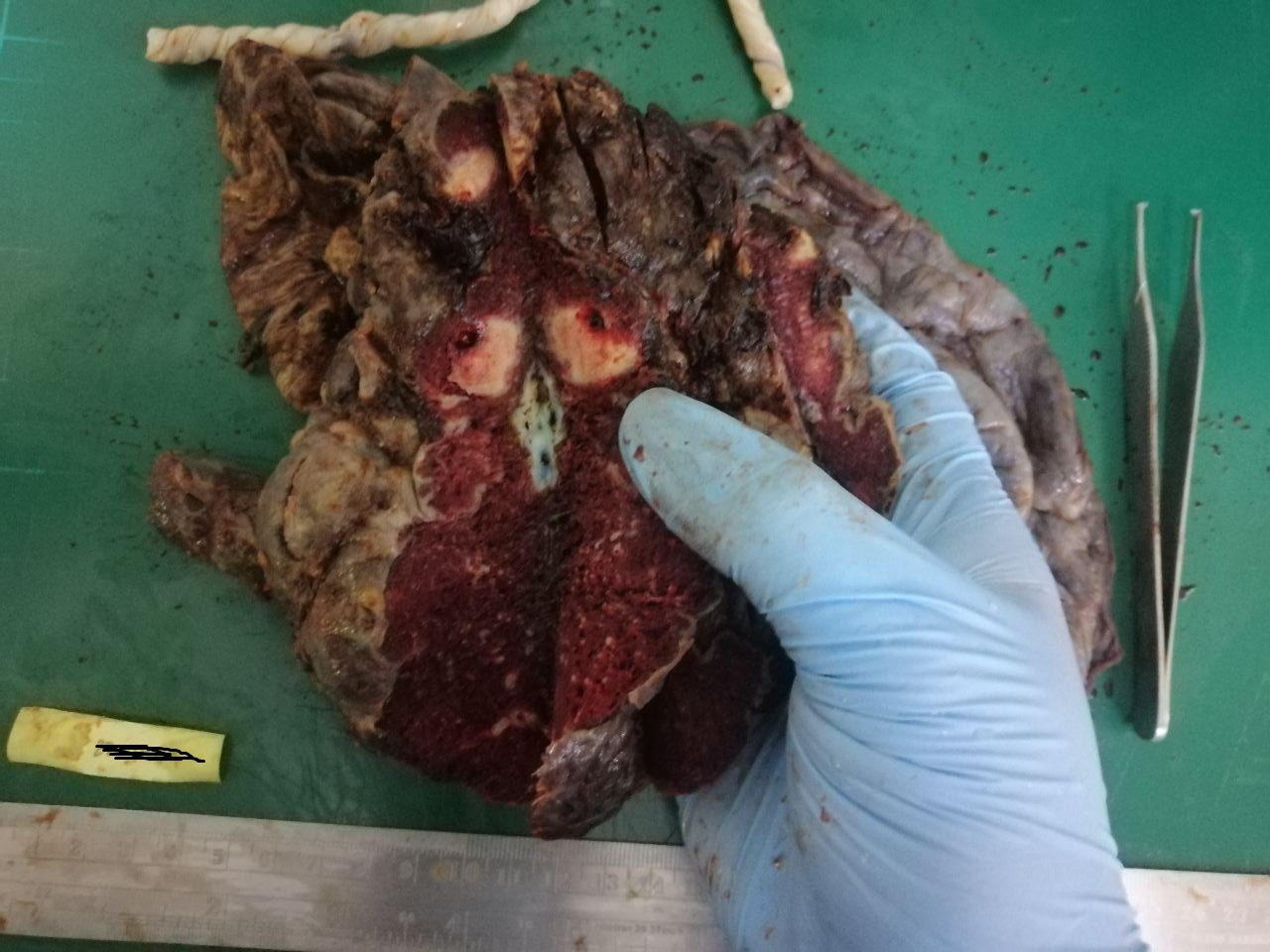
Following up with the case I shared about a week ago, these are the slides that came out from those placental nodules. The first images are some sections taken from the grossly normal parts of the specimen.
Taken at scanner view (40x magnification)
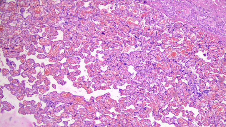
You can see several vari-sized chorionic villi lined by trophoblasts. You’ll also see these villi have some capillaries within their stroma. Even if no one tells me the age of gestation, seeing vascular villi hints they are on their second to third trimester as a sign of maturity. They need to have capillaries proliferating to support the growing fetus.
Taken at low power view (100x magnification)
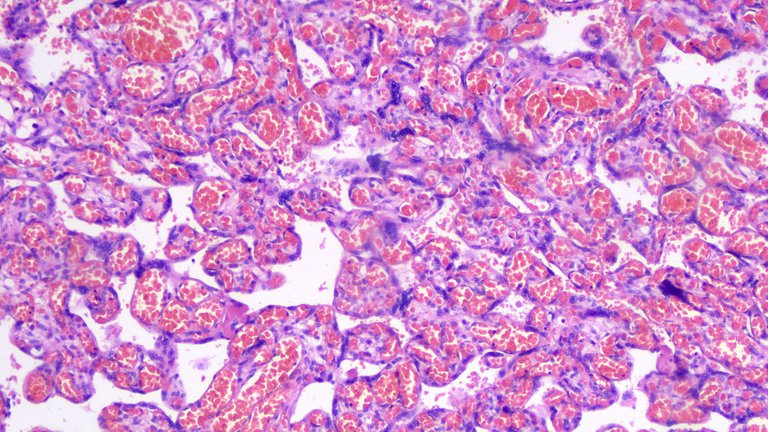
You’ll see a predominance of small to medium sized villi which is another indicator that this is a mature placenta. I would be looking for vascularity, size of villi, trophoblastic proliferation, and trophoblasts lining the villi as clues to date the placental age. But from experience, the most reliable indicators are vascularity and size.
Taken at high power view (400x magnification)
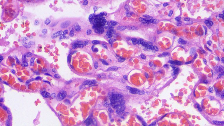
Just emphasizing how vascular villi can be especially during the trimester. This case was taken from a placenta dated on their early term labor.
Now for the nodules identified.
Taken at scanner view (40x magnification)
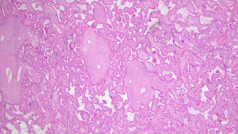
You can see everything is predominantly pink or hyalinized indicating it’s all dead tissues.
Taken at low power view (100x magnification)
There’s a lack of basophilic nuclei from viable cells and notice the lack of red blood cells indicating no vascular supply in contrast to the previous sections of normal placenta.
Taken at high power view (400x magnification)

The blue parts are just pigments from the stains used. This supports my gross diagnosis that those nodules present were old infarcts probably happened weeks prior to the actual labor. This limited some blood supply to the fetus but fortunately the baby lived and delivered via C-section.
If you made it this far reading, thank you for your time.
Posted with STEMGeeks
Congratulations @adamada.stem! You have completed the following achievement on the Hive blockchain and have been rewarded with new badge(s):
Your next target is to reach 8000 upvotes.
You can view your badges on your board and compare yourself to others in the Ranking
If you no longer want to receive notifications, reply to this comment with the word
STOPCheck out the last post from @hivebuzz:
Support the HiveBuzz project. Vote for our proposal!