Had my First Case of Conventional Osteosarcoma
Probably one of the most graphic images I've shared so far. Behold, a leg cut in half. This was bathed in formalin, that's why it turned into the color it is.
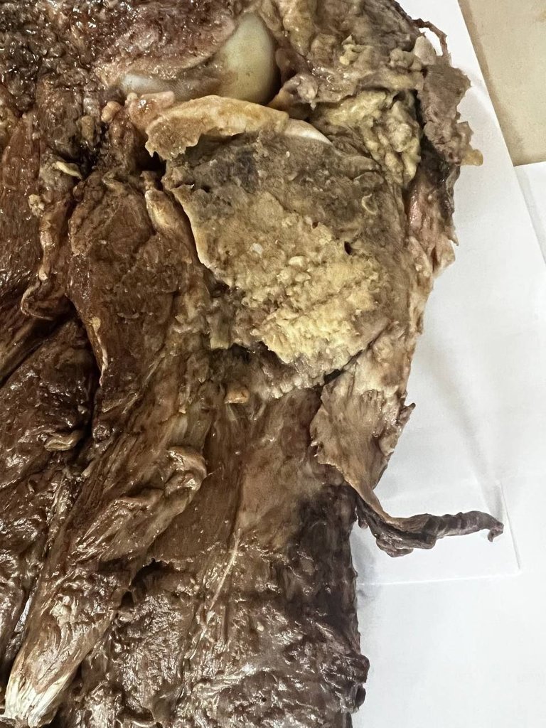
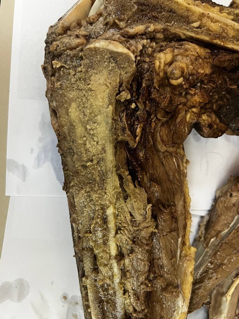
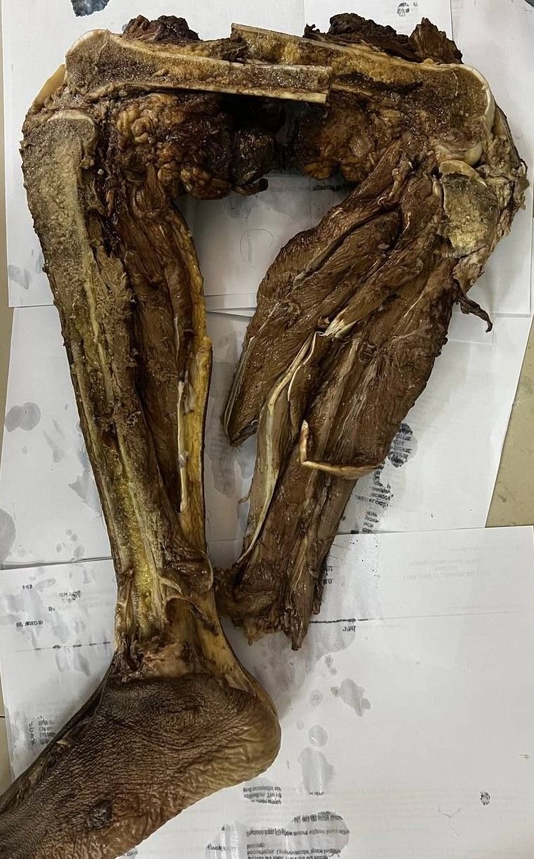
It's a leg of a 14-year old male treated with chemotherapy prior to amputation. He was a diagnosed case of Conventional Osteosarcoma, Fibroblastic Type with a widest tumor dimension of 8cm just confined to the proximal tibia.
There are other variants of Osteosarcoma and the cancer itself is common on the younger age group where growth spurts happen which becomes an enticing place to grow as a tumor. Now part of the risk is genetic predisposition but we won't go into those extensive details here.
Dealing with post chemo specimens adds an extra layer of work when it comes to sectioning. Imagine taking a slab of the tumor and then separating the areas with an imaginary tables just to map out the extent of tumor involvement.
The purpose is to determine the extent and whether the chemotherapy killed the tumor cells. Naturally, if more of the tumor cells got killed it spells out good prognosis but this is also influenced by the extent of involvement whether the tumor has already spread to other parts of the body.
I forgot to take photomicrographs of the specimen as it wasn't really on my head to make some content out of each specimen I process. To compensate, I found an exact image of what I saw under the microscope from here.
I haven't been cutting specimens due to my post moved to the clinical pathology where I'm mostly expected to be on the laboratory or doing miscellaneous work. I was lucky enough to get this one done because I needed the practice. I'm sure I'll know pain once I'm back on the histopath department.
If you made it this far reading, thank you for your time.
Posted with STEMGeeks
I had a type of soft tissue sarcoma that was also child Hood related but got it when I was 29!
Double chemo + radiation and stage four "terminal" spread to various places mostly muscle but also spine...
now 4 yrs later its dead not gone in the spine, steel hip, reinforced both upper leg bones, biggest tumor was around 15cm.
Misdiagnosed as sciatica till i felt bumps in my shoulder /back
Fully recovered running up mountains, renovating houses etc
It's a bimodal age distribution from early teens and 40s. Some cases can happened in between the age ranges and they tend to be aggressive. Glad you survived that, and damn, 15cm? that's huge.
Did you guys use a dunking tank with formalin or something?
And dang, an entire leg removed for a tumor in the tibia...almost seems excessive, but I'm not a physician.
Those large plastic boxes for storage were filled with formalin for this. It's an aggressive tumor and metastasis was often found during the time of diagnosis or occult spread already happened. Though this case had all the margins negative for tumor, it doesn't rule out that some of it has spread already but not clinically detectable yet.
That’s rough for the kid. Poor him.
Damn, that's grotesque and sad to lose a leg at 14
Better lost than be a source of more trouble. The chemo was to limit the growth and potential spread. Unless the spread has been extensive already, resection with chemo was the staple choice.
Congratulations @adamada.stem! You have completed the following achievement on the Hive blockchain and have been rewarded with new badge(s):
Your next target is to reach 6000 upvotes.
You can view your badges on your board and compare yourself to others in the Ranking
If you no longer want to receive notifications, reply to this comment with the word
STOPCheck out the last post from @hivebuzz:
Support the HiveBuzz project. Vote for our proposal!