Followed up on those immunohistochemical stains TTF-1 and Calretinin
This is a follow up post on the previous case I shared. I got the images for the immunohistochemical stains from that pleural fluid sample.
Short Recap:
The specimen sent a liter of pleural fluid for staining and interpretation. The clinicians were entertaining malignancy versus an infectious process. So we found the clusters of cells below.
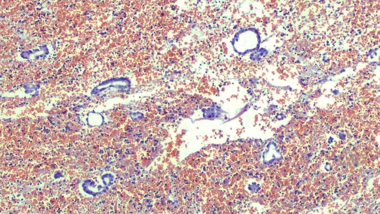
Now these look suspicious for malignancy. The next step is determining whether these were mesothelial cells that are just reactive to inflammation or actual atypical cells coming from the lungs.
Immunohistochemical stains help confirm the diagnosis by staining cells that have unknown origin. It’s an understatement to what they can potentially do in practice but expounding on it further is a bit of information overload for those not into immunology and fields alike. At the simplest way to put it, if the cells are stained using stains that stain for X tissue, then there’s a high chance they are indeed X tissue.
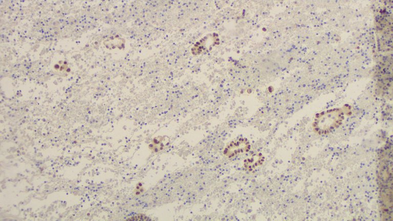
Some cancer cells look so bizarre that it’s impossible to determine which part of the body they came from. So the method to clinch the diagnosis is the use of stains that help narrow down where the source is. On this this, the stains used were Thyroid Transcription Factor-1 and Calretinin.
If Calretinin stains turned out positive (evidenced by brown pigmentation on nuclear and cytoplasm), the atypical cells are most likely just reactive mesothelial cells leaning on a benign reaction to inflammation but can’t totally rule out malignancy.
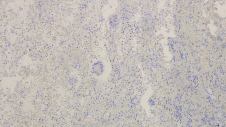
It didn't stain.
If TTF-1 stains turned out positive (evidenced by nuclear staining), the atypical cells would point to a malignant source from the lungs. Now this isn’t always the case but more about it later.
As you can see, TTF-1 stained positive.
Now it’s not all the time a tumor will respond to a stain as intended as there are variants that don’t but that’s another can of worms to talk about. Immunohistochemical stains help eliminate other differentials but these aren’t used as stand-alone tests for diagnosing. A single stain can also be positive for at least 3 organs so another stain is usually used in combination to help narrow down the source (ideally in practice)
TTF-1 can also stain thyroid cells, so if the clinicians suspect a thyroid or lung origin, another set of IHC stains will be used but on this case, the patient already has a lung mass so no further workup was done on locating the source.
If you made it this far reading, thank you for your time.
Posted with STEMGeeks