Atypical Endometriosis, an Incidental Finding
This was taken from an endometriotic cyst (ovary containing ectopic endometrial glands and stroma). Endometriosis is histologically apparent when there are endometrial glands and stroma on areas outside the uterine endometrium. These can spread to proximal areas within the female genital tract up to unconventional distant areas. There are some theories explaining how one can find ectopic endometrial tissues outside the endometrium but that’s not the focus on this post.
I didn’t take a picture when I was grossing this specimen as endometriotic cysts were common. But if for reference, the ovary looked something like this.
Endometrial tissues implanted on parts where they aren’t supposed to be also react to hormonal stimulation. So if it’s that time of the month to bleed, these sites may bleed too. This explains the chocolate looking contents as these are blood pools with degenerated RBCs due to it being a chronic condition.
Taken at scanner view (40x magnification)
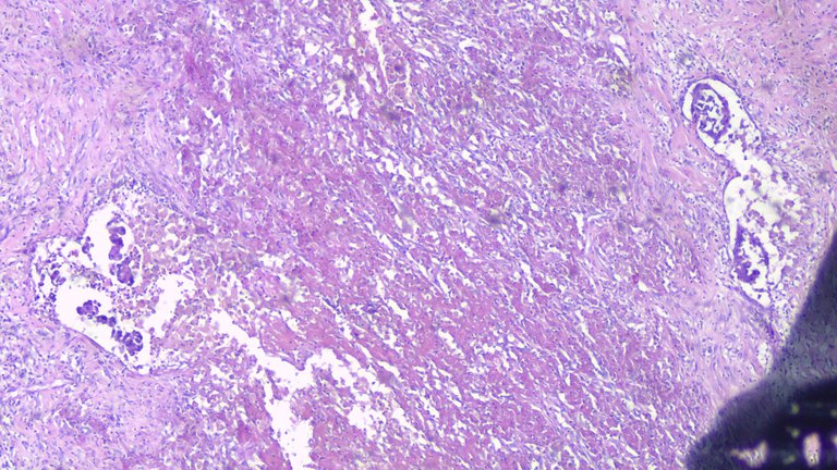
Clusters of endometrial glands surrounded by sheets of foamy and hemosiderin-laden macrophages. You would expect to see hemosiderin-laden macrophages on these cases as a staple finding because chronic bleeding. These type of macrophages recycle iron from broken down red blood cells and take up the brown pigments. The surrounding areas look necrotic because of how long the condition has deteriorated the lining and surrounding tissues from repeated hemorrhage.
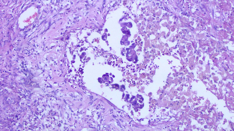
Taken at low power view (100x magnification)
Taken at high power view (400x magnification)
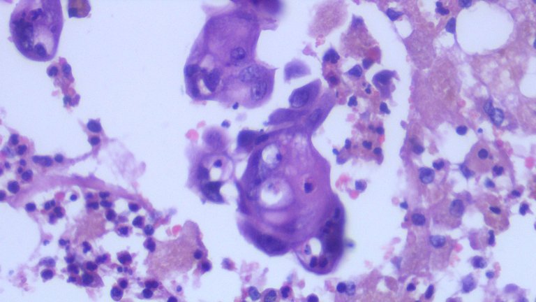
Seen as minute clusters of hyperchromatic to vesicular, round to ovoid nuclei with inconspicuous to prominent nucleoli and abundant eosinophilic cytoplasm.
This was an incidental finding and it happens rarely. It’s the first time I encountered such case. The impact of this diagnosis is how atypical endometriosis raises the risk of endometrioid ovarian carcinoma as it’s like a precursor lesion.
Sometimes endometriotic cysts don’t really have that classic textbook picture where you’ll see endometrial glands and stroma well preserved. If the patient has this condition for a long time, I’d usually see only sheets of hemosiderin-laden macrophages lining the cyst and several foci of hemorrhage. During this time, it can be called hemorrhagic cyst consistent with endometriotic cyst.
As a general rule, histologic diagnosis requires the element present under the microscope to support the diagnosis but some cases where there’s degeneration of the endometrial glands and stroma due to the conditions being chronic, it’s hard to demonstrate it on sections. But if one correlates it with the clinical condition, grossing, and microscopic findings, this usually makes it up.
If you made it this far reading, thank you for your time.
Posted with STEMGeeks
I read that men can get endometriosis too although it's very rare. Weird disease
Not exactly the type of information I'd encounter when discussing the subject but a quick look up makes it possible to have those due to prolonged therapy. I learned something new on this comment. I'd be giddy if I get to have that case. The other possibility is seeing these cases occur more in the future due to how relative the "gender" tagging can be, like some identifying as men but not biologically men.
Yes. Apparently liver cirrhosis and any other condition increasing estrogen in men can cause it too
Interesting case of endometryosis ! I guess nature is tricky sometimes
!1UP
You have received a 1UP from @gwajnberg!
@stem-curator, @vyb-curator, @pob-curator, @neoxag-curator, @cent-curator
And they will bring !PIZZA 🍕.
Learn more about our delegation service to earn daily rewards. Join the Cartel on Discord.
PIZZA Holders sent $PIZZA tips in this post's comments:
@curation-cartel(5/20) tipped @adamada.stem (x1)
You can now send $PIZZA tips in Discord via tip.cc!
Interesting content! Next time consider adding some sources so one can learn more about this whole thing :)
Thank you for stopping by. I intentionally make it as simple as possible to understand without being too textbook looking. The things I mentioned here can be googled easily of one wants to compare it to what the textbook says. Think of the post as a retell of what I see in practice told in a manner where people dont have to think hard if I am talking about apples and how it is a fruit.
But if there is a specific detail you need, I'll look it up again. The reference I used are mostly from a paid subscription of an online textbook by WHO and a physical copy of a textbook, on this case published by Blaustein. So even linking those may mean having to go through a paywall or finding a soft copy and rummaging for the snippet of info on the pdf file. Not really a reader friendly experience.
As for the images, it's taken from what I saw on the microscope so I don't mention the sources that I own. Images I don't have sources on them.
Got it, but still it is a curation standard to some, you know. It also could make your post more classy :)
I debated about presentation when I started posting stuff like this in the past. I don't disagree with the input here as I see it having sense to present information in a classy way. But I still opted to just do things this way on the basis where I'm sharing stuff in chill mode and not trying to impress a medical community which requires an intensive amount of detail. Going that route removes my motivation to even post.
I think about the general reader experience. If a random stranger, none medically inclined came upon my post, information should be presented in a way that it's easy to grasp intuitively without a medical background and if there are any jargons or concepts present, these can be easily Googled up.
It's the same pet peeve I have with thesis type of writing, how so something so interesting can bore me when I have to be fed with terms that can be simplified and this just prevents people from appreciating the information and science behind. But this is just my preference.
Gotcha. You don't have to add "an intensive amount of detail", feed people with complicated terms or write a thesis to bore other people and yourself. Adding two or three links, though won't hurt anyone and it could actually help you.
Yeah, I'm a bit hesitant to link people to https://tumourclassification.iarc.who.int/welcome/ where I got the info and be greeted with a paywall to know. There is also - Blaustein. But that's where I got my sources from. The point is being considerate with not putting links that really give the reader answers they want if they have to go through different hoops to get it.
If there is a specific part on the post one wants to verify, I can backread it and give the answer and that gives me an idea that people actually read the post.
Hehe, so you want to be "considerate" of the reader. Thinking too much about how to evade the little suggestion can lead you to use ad hoc arguments ;)
I am not evading the suggestion. I get the suggestion. I already told you why I dont want follow the suggestion. If there is a piece of information that isn't clear, let me know so that I can double back on that detail and expound it so that I know you actually read it and interested. This is the voice I want my post to have.
I peeked into your post with that comment because I was trying to be nice and politically correct by hinting to you that adding some sources in your post might help the work of some curators and help you get support.
I understand your self-determination and self-confidence to do the post the way you want.
The fact that you used the word trying and politically correct implies a contrast to what you were sincerely thinking.
It's a fair point and I took your initial points of contact as constructive criticism. The parts where it started to get annoying was drilling on the subject further. I get the part where you are pointing me to the right direction to get those curation incentives for sharing, I really do. But it became frustrating to explain that you assumed I didn't and disregarded why I insist on doing things my way despite being frank why I don't subscribe to the curation norms.
I'm not invested in the conversation as it's all a matter of preference how one frames their post and curator bias if they think the post is worthy of support. But I do appreciate you taking the time to visit. My confidence comes from doing the actual work and living the profession that is privileged to share this information.
I don't see how that conclusion follows those premises. Even if I was thinking something different, the set of my possible thoughts is too broad and could mean many things.
This is my point (I hope I can clarify it finally). Often, when you are too direct or honest, some people get offended. Trying to be nicer by rephrasing what you say is called "euphemism", which in turn means "trying to be politically correct". It is "trying" because there is no guarantee that it will work in some cases and people will still be offended by the expression used. For example:
"You made a tactically unsound move" is code for "you blundered" (typical in chess).
"Person of short stature" is code for "dwarf".
"Next time consider adding some sources so one can learn more about all this :)" was my failed euphemistic attempt to say "Next time add sources because I shouldn't cure/vote you if you don't meet some criteria".
Then you started a series of lengthy responses that included several points beyond "adding two or three links". It suggested to me that you were a bit defensive, but my intent was not to criticize or offend. I understand that you didn't get it the euphemism. Perhaps more contextual knowledge was needed.
Ok, I know what your position is and I hope I have clarified the reasons after my visit and comments. We are done :)
I don't think nice people need to try to be nice to fit the label of being nice but that's just my opinion.
I understood the nudge to source the information I post properly and felt the need to elaborate why I opted not to because my preference in framing my post does not conform to the curation criteria curators may have set for their respective communities.
I already made my peace with the issue prior to posting a series of STEM related topics months back. If ever I post information that is unclear, I am open to discuss as this is the risk of posting without sources.
For future reference, it's all on WHO most of the time but one can take also use pathologyoutlines as this page contains summaries from WHO and other sources of literature to topics I talk about.
Shame...would have loved to see the afflicted organs.
!discovery 37
Endometriosis can afflict odd places like knees and colon. I had a case where they resected an entire right colon cause it presented as an obstruction and looked like cancer. They look the same on imaging as any other cancer and tuberculosis. Most are managed as hormonal therapy but resection usually does it, there is still a chance for recurrence though.
What makes this case rare isnt the endometriotic cyst part which ia common but the atypical epithelium that transformed associated with it.
This post was shared and voted inside the discord by the curators team of discovery-it
Join our community! hive-193212
Discovery-it is also a Witness, vote for us here
Delegate to us for passive income. Check our 80% fee-back Program
Your content has been voted as a part of Encouragement program. Keep up the good work!
Use Ecency daily to boost your growth on platform!
Support Ecency
Vote for new Proposal
Delegate HP and earn more