A case of Melanoma
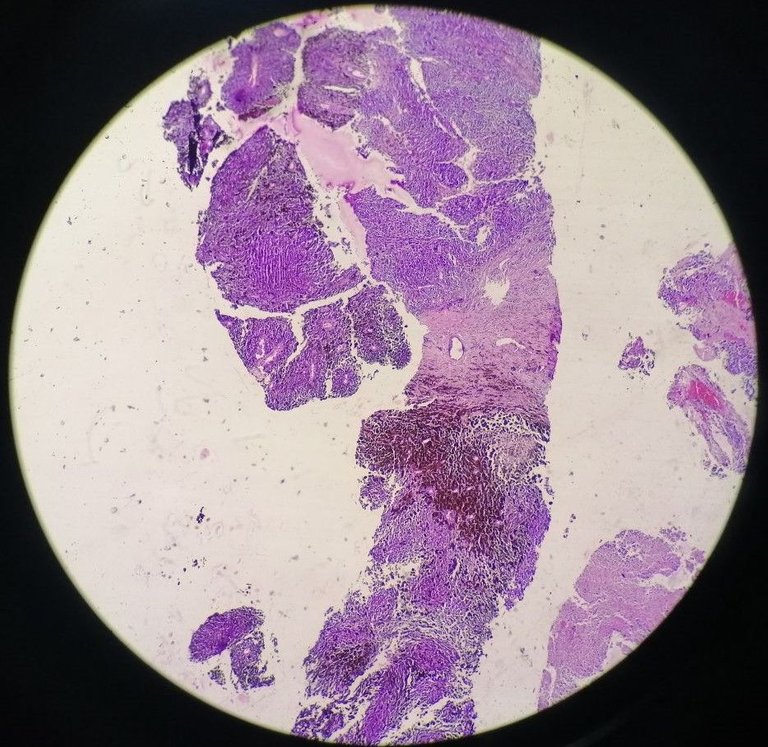
A 44-year old male with a right thigh mass. I only received the specimen so I never saw what it really looked like. This is normal but ideally the ones sending the request should have attached a picture of the lesion. They sent around pinched sized tissue fragments.
Anyway, this was an easy case as soon as I saw the scanner view. It's that brown to black pigment surrounded by malignant looking cells that gave the case away.
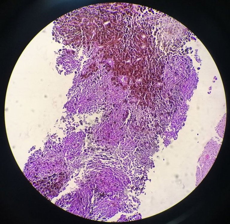
Melanocytes are the cells responsible for producing the melanin pigment which gives the skin its natural color. If you find them at the deepest layers or other parts where they shouldn't be, that's likely malignant.
One way to get confused them for other pigment holding cells are your hemosiderophages which are your macrophage (member of the white blood cells that taken residence to tissues). These take up hemosiderin, an iron released from dead red blood cells and they take these up for recycling the iron.
There's no hemorrhage surrounding the area and the malignant looking cells seen below surrounding the pigmented ones gives it away. It's hard to tell between the two if they happen to be in singles. There are also cases where melanoma may not have any pigments at all which makes the diagnosis difficult and more reliant on immunohistochemical stains to confirm.
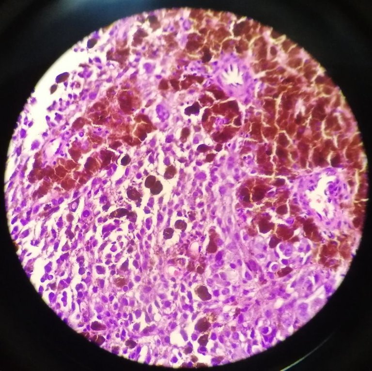
We still do immunohistochemical stains like S100 and HMB-45 for confirmation but it's left for the clinician to pursue those. It's like, yeah morphologically it looks like what we think it is but there's always room for error to entertain so stains are suggested just in case.
Melanoma is an aggressive tumor where skip metastasis is common. So if by chance that it gets diagnosed at the skin, it's not uncommon to have incidental findings on other organs after thorough workup.
If you made it this far reading, thank you for your time.
Posted with STEMGeeks
How much time does it take for you to do one slide?
Depends on the specimen, if it's cytology like pap smears and other fluids, may take more than 15 min. If it's something as benign as appendicitis, less than 5 mins.
I can't really gauge the time but it's more along the lines of seeing it many times subjectively makes me feel the time flies faster. I know some pap smears take less than 5 to 10 minutes. Some institutions would have a cytotechnician to do those reading but ours don't so we get swamped from those on top of the tissue biopsies.
The longest slides are those from rare specimens like brain tissue, salivary, and pancreas. Other than reading the slide, one needs extra reading from the current guidelines to diagnose which has their own specifications and it's easy to get lost track per organ.