Se definen como las alteraciones estructurales del corazón o de los grandes vasos. Son las malformaciones congénitas más comunes y se producen durante el desarrollo embrionario del corazón, aproximadamente entre la semana 3 y semana 10 de gestación.
They are defined as structural alterations of the heart or great vessels. They are the most common congenital malformations and occur during the embryonic development of the heart, approximately between week 3 and week 10 of gestation.
No hay una causa en específico, de hecho, la mayoría de las cardiopatías congénitas se presentan sin ninguna causa aparente, pero, se pueden mencionar algunas cusas que pudieran motivar la aparición de la misma, como por ejemplo: factores genéticos, uso de fármacos teratógenos durante el embarazo, abuso de alcohol y/o drogas durante el embarazo, infecciones maternas, etc.
There is no specific cause, in fact, most congenital heart disease occurs without any apparent cause, but some causes that could lead to its appearance can be mentioned, such as: genetic factors, use of teratogenic drugs during pregnancy, alcohol and/or drug abuse during pregnancy, maternal infections, etc.

Fuente / Source

¿CÓMO SE CLASIFICAN Y CUÁLES SON LAS MÁS FRECUENTES?
¿HOW ARE THEY CLASSIFIED AND WHICH ARE THE MOST FREQUENT?
Éstas se dividen en: cianógenas y acianógenas.
- Cianógenas: la sangre desoxigenada no sufre el proceso de oxigenación normal, y, por ende, la circulación sistémica está repleta de sangre desoxigenada.
- Acianógena: la circulación sistémica está comandada por sangre oxigenada, que es lo que normalmente debería suceder.
Las cardiopatías congénitas más frecuentes son:
| CIANÓGENAS | ACIANÓGENAS |
|---|
| Tetralogía de Fallot | Comunicación interventricular |
| Transposición de grandes vasos | Comunicación interauricular |
| Atresia tricuspídea | Conducto arterioso persistente |
| Coartación aórtica |
| Estenosis aórtica |
| Estenosis pulmonar |
NOTA: La cianosis es la coloración azulada de piel y mucosa, se observa principalmente en extremidades, lóbulo de la oreja, punta de la nariz, labios, lengua, etc.
These are divided into: cyanogens and acyanogens.
- Cyanogenic: deoxygenated blood does not undergo the normal oxygenation process, and, therefore, the systemic circulation is full of deoxygenated blood.
- Acyanogenic: the systemic circulation is commanded by oxygenated blood, which is what should normally happen.
The most common congenital heart diseases are:
| CYANOGENES | ACYANOGENS |
|---|
| Tetralogy of Fallot | Ventricular septal defect |
| Transposition of the great vessels | Atrial communication |
| Tricuspid atresia | Patent ductus arteriosus |
| Aortic coarctation |
| Aortic stenosis |
| Pulmonary stenosis |
NOTE: Cyanosis is the bluish discoloration of the skin and mucosa, it is observed mainly in the extremities, earlobe, tip of the nose, lips, tongue, etc.
CARDIOPATÍAS CONGÉNITAS CIANÓGENAS
CYANOGENIC CONGENITAL HEART DISEASE
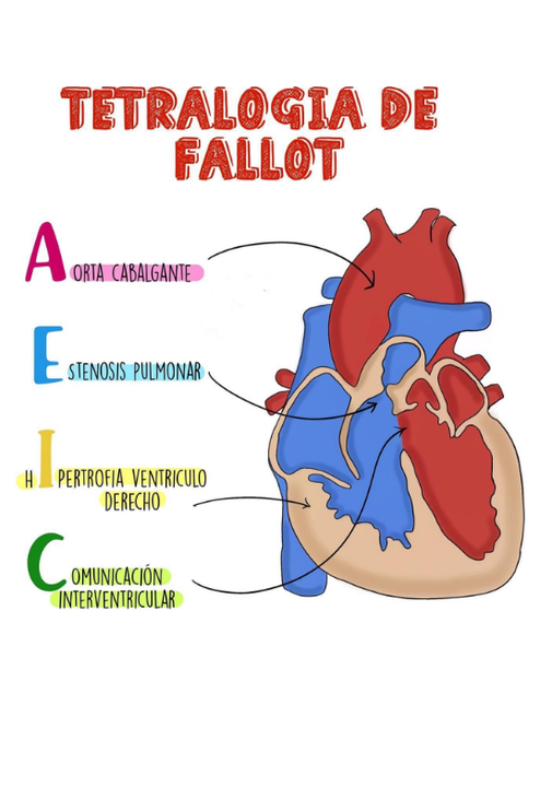
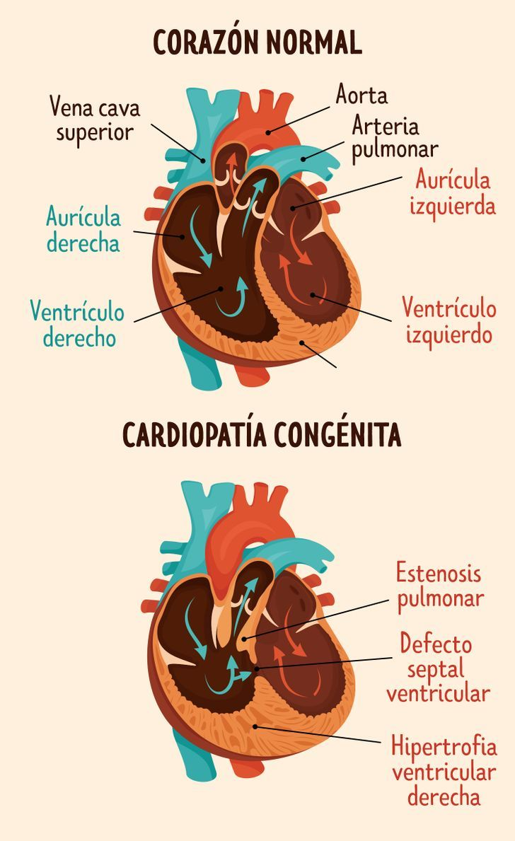
1. Tetralogía de Fallot:
Se caracteriza por presentar 4 anomalíaas: hipertrofia ventricular derecha, comunicación interventricular, estenosis pulmunar y cabalgamiento de la aorta.
1. Tetralogy of Fallot:
It is characterized by presenting 4 anomalies: right ventricular hypertrophy, interventricular communication, pulmonary stenosis and overriding of the aorta.
Fuente / Source
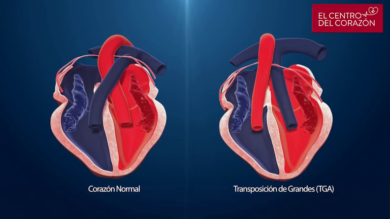
2. Transposición de grandes vasos:
Es una patología incompatible con la vida. Lo normal es que la arteria pulmonar salga del ventrículo derecho y que la arteria aorta salga del ventrículo izquierdo, en esta patología, es al revés; esto crea dos sistemas circulatorios totalmente independientes, lo que provoca la muerte del recién nacido si no se corrige de manera inmediata.
2. Transposition of the great vessels:
It is a pathology incompatible with life. The normal thing is that the pulmonary artery leaves the right ventricle and that the aorta artery leaves the left ventricle, in this pathology, it is the other way around; this creates two completely independent circulatory systems, leading to the death of the newborn if not corrected immediately.
Fuente / Source
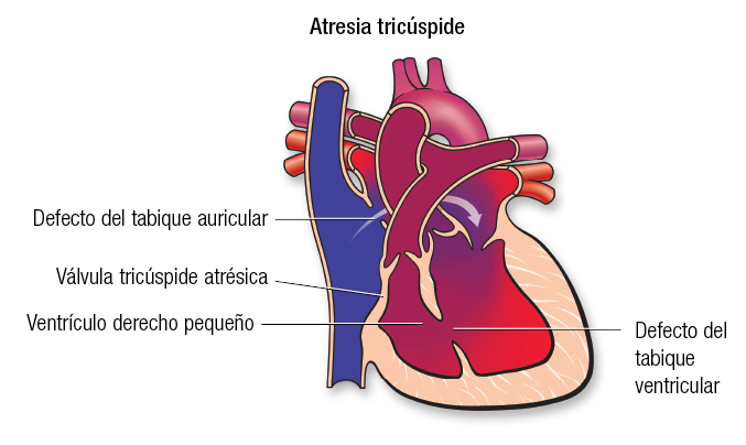
3. Atresia tricuspídea:
Se debe a la ausencia de la válvula tricuspídea; que es la encargada de comunicar a la aurícula derecha con el ventrículo derecho, por tal motivo, se presentan otros defectos para contrarrestar el defecto principal, pero, la sangre que llega al ventrículo derecho no puede ser bombeada de manera eficiente hacia los pulmones a través de la arteria pulmonar.
3. Tricuspid atresia:
It is due to the absence of the tricuspid valve; which is responsible for communicating the right atrium with the right ventricle, for this reason, other defects are presented to counteract the main defect, but the blood that reaches the right ventricle cannot be pumped efficiently to the lungs through of the pulmonary artery.

Fuente / Source
CARDIOPATÍAS CONGÉNITAS ACIANÓGENAS
ACYANOGENIC CONGENITAL HEART DISEASE
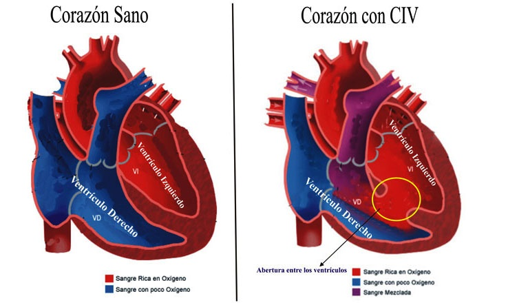
1. Comunicación interventricular:
Se debe a la presencia de uno o varios orificios en el tabique que separa el ventrículo derecho del izquierdo, lo cual permite la comunicación entre ambos.
1. Ventricular septal defect:
It is due to the presence of one or more holes in the septum that separates the right ventricle from the left, which allows communication between the two.
Fuente / Source
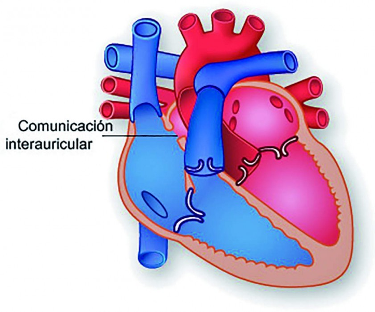
2. Comunicación interauricular:
Aquí, la presencia de los orificios se encuentra a nivel del tabique que separa ambas aurículas del corazón.
2. Atrial communication:
Here, the presence of the holes is at the level of the septum that separates both atria from the heart.
Fuente / Source
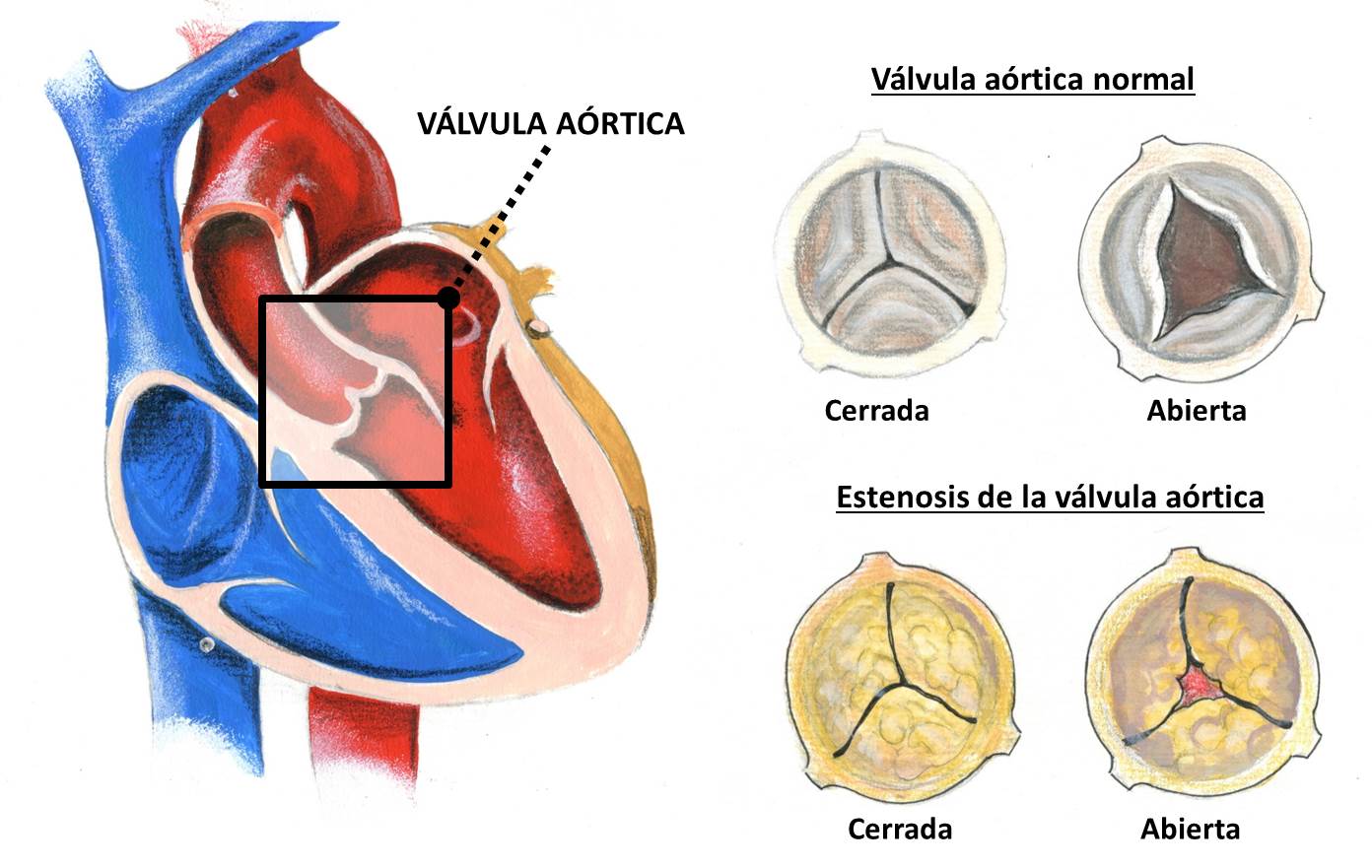
3. Estenosis aórtica:
La arteria aorta (que distribuye la sangre al resto del cuerpo), en su salida desde el corazón, tiene una válvula denominada: válvula aórtica; en esta patología, la válvula aórtica presenta una alteración, lo que produce una obstrucción de la salida de la sangre desde el ventrículo izquierdo.
3. Aortic stenosis:
The aortic artery (which distributes blood to the rest of the body), at its outlet from the heart, has a valve called: aortic valve; In this pathology, the aortic valve presents an alteration, which produces an obstruction of the outflow of blood from the left ventricle.

Fuente / Source
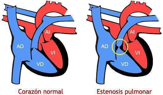
4. Estenosis pulmonar:
Aquí el defecto se encuentra en la arteria pulmunar, la cual sale desde el ventrículo derecho y distribuye la sangre hacia los pulmones, para que pueda ser oxigenada.
4. Pulmonary stenosis:
Here the defect is in the pulmonary artery, which comes out of the right ventricle and distributes the blood to the lungs, so that it can be oxygenated.

Fuente / Source
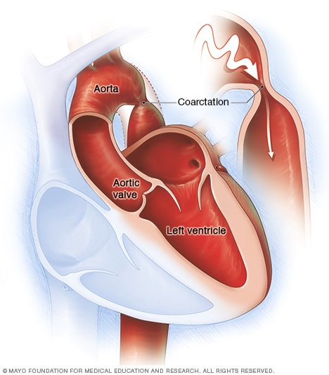
5. Coartación aórtica:
Se debe al estrechamiento de una porción de la arteria aorta, lo que impide que la sangre de distribuya de manera eficiente al resto del organismo.
5. Aortic coarctation:
It is due to the narrowing of a portion of the aortic artery, which prevents blood from being distributed efficiently to the rest of the body.
Fuente / Source
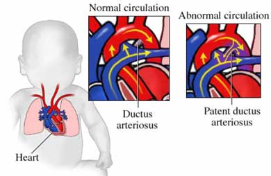
6. Conducto arterioso persistente:
Durante la vida intrauterina, existe una comunicación entre la arteria aorta y la arteria pulmonar, la misma debe cerrarse luego del nacimiento. Esto dará lugar al llamado: ligamento arterioso; que pasará a ser la estructura definitiva en dicho lugar. Si esto no ocurre, se produce un aumento de volumen y presión tanto en la aurícula izquierda como en el ventrículo izquierdo, que se puede comportar como una insuficiencia cardíaca y edema pulmonar.
6. Patent ductus arteriosus:
During intrauterine life, there is a communication between the aorta and the pulmonary artery, which must be closed after birth. This will give rise to the so-called: ligamentum arteriosus; which will become the definitive structure in said place. If this does not happen, there is an increase in volume and pressure in both the left atrium and the left ventricle, which can behave as heart failure and pulmonary edema.
Fuente / Source
¿ES POSIBLE DETECTAR CARDIOPATÍAS CONGÉNITAS A TIEMPO?
¿IS IT POSSIBLE TO DETECT CONGENITAL HEART DISEASE EARLY?
Sí, aunque existen ciertas cardiopatías que no se pueden diagnosticar prenatalmente, motivado a esto, existen una serie de signos y síntomas que pueden ayudar a detectarla en los primeros meses de vida.
La ecografía fetal es un método muy utilizado para diagnosticar alteraciones a nivel cardíaco, esto puede ser una gran ventaja para actuar tan pronto sea necesario.
Dentro de los signos y síntomas que podemos encontrar en un recién nacido con cardiopatía congénita, tenemos: cianosis, frecuencia cardíaca aumentada, alteraciones a nivel de los pulsos, dificultad para respirar, entre otros.
Yes, although there are certain heart diseases that cannot be diagnosed prenatally, motivated by this, there are a series of signs and symptoms that can help detect it in the first months of life.
Fetal ultrasound is a method widely used to diagnose alterations at the cardiac level, this can be a great advantage to act as soon as necessary.
Among the signs and symptoms that we can find in a newborn with congenital heart disease, we have: cyanosis, increased heart rate, alterations at the pulse level, difficulty breathing, among others.
Espero haya sido de tu agrado el contenido de éste post, agradezco su colaboración con comentarios, upvote's y repost. Hasta la próxima.
I hope the content of this post has been to your liking, I appreciate your collaboration with comments, upvote's and repost. Until next time.


PRIMUM NON NOCERE


/Telegram_(software)-X-Black-Logo.wine.png)
/Discord_(software)-Logo-Black-Logo.wine.png)
https://twitter.com/gablejandrovv/status/1493211644180287489
The rewards earned on this comment will go directly to the person sharing the post on Twitter as long as they are registered with @poshtoken. Sign up at https://hiveposh.com.