Mammalian Tissue Under the Microscope
Hello everyone. I'm back with more images taken with my new microscope. For this this post, I thought that I would share some images of mammalian tissue samples from my collection of permanent slides.
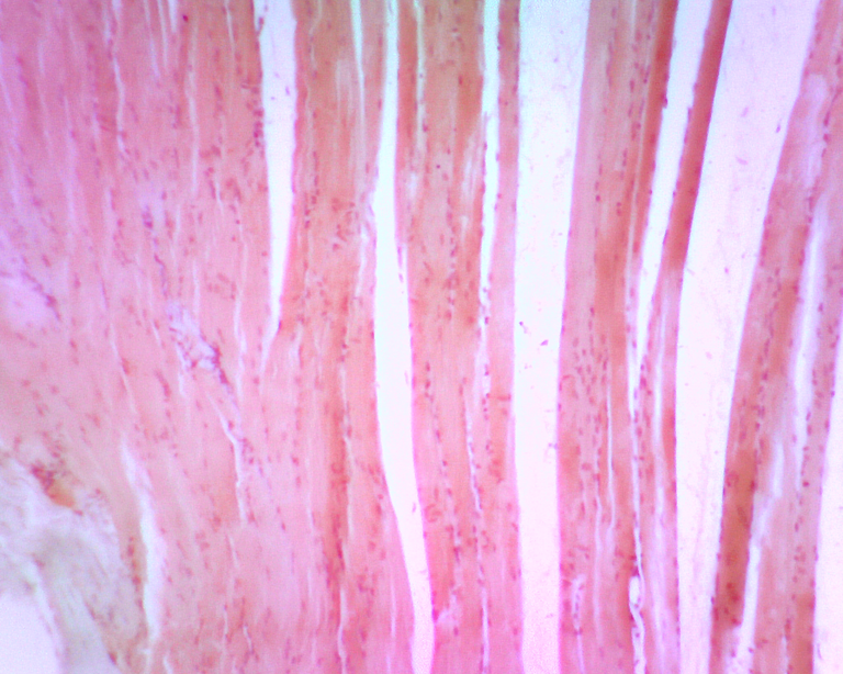
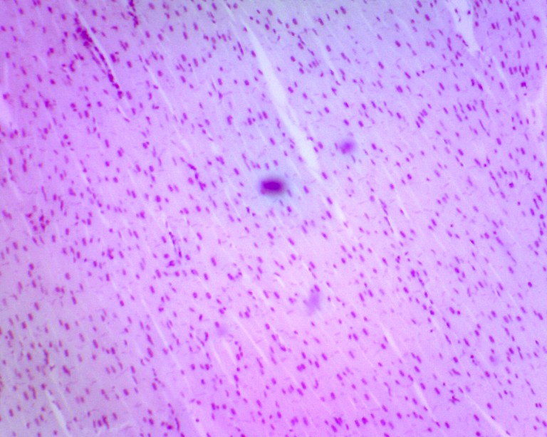
Striated Muscle:
Striated muscle is tissue comprised of functional units known as sacromeres and is subdivided into two categories: cardiac muscle and skeletal muscle. Striated muscle differs from smooth muscle in that striated muscle fibers are cylindrical in shape and, with the exception of cardiac muscle, connect to some component of the skeletal system. Smooth muscle fibers on the other hand are more spindle-like in structure and make up muscle tissue not connected to the skeletal system such as internal organs and the intestines.
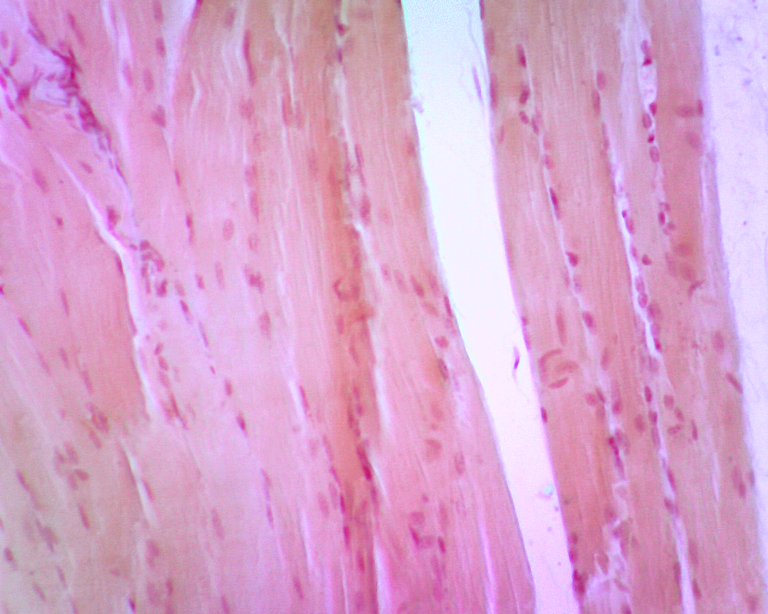
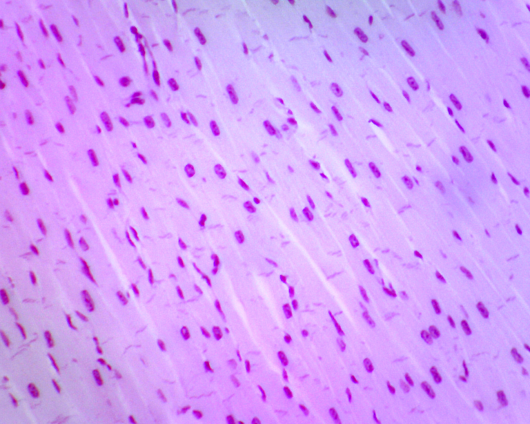
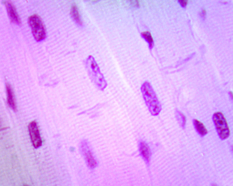
Cardiac Muscle:
Cardiac muscle is a form of striated muscle that comprises the tissue that makes up the heart. Unlike skeletal muscles, whose contraction can be consciously controlled, the involuntary contraction and release of cardiac muscle fibers in the heart is regulated by specialized cells known as pacemaker cells.
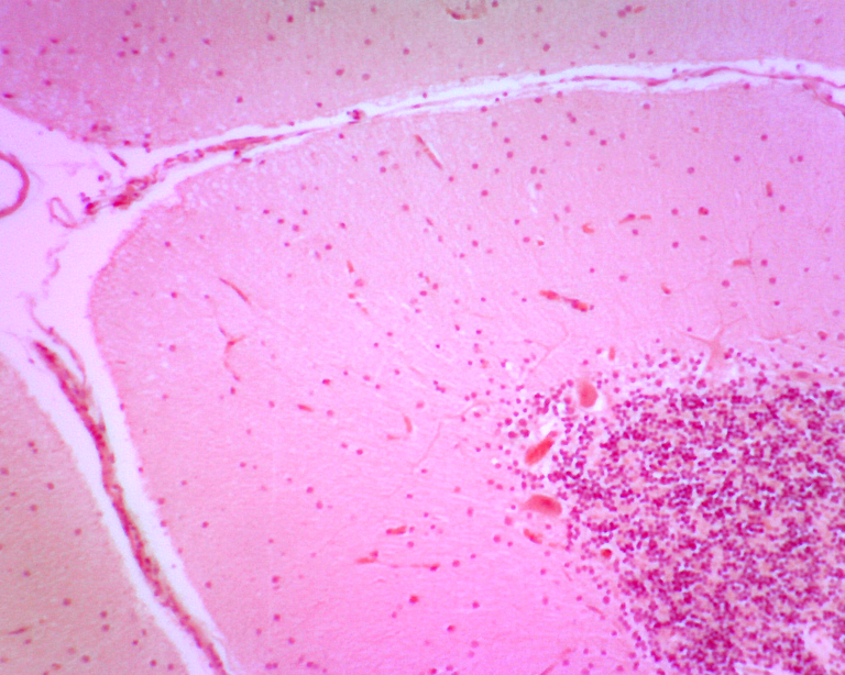
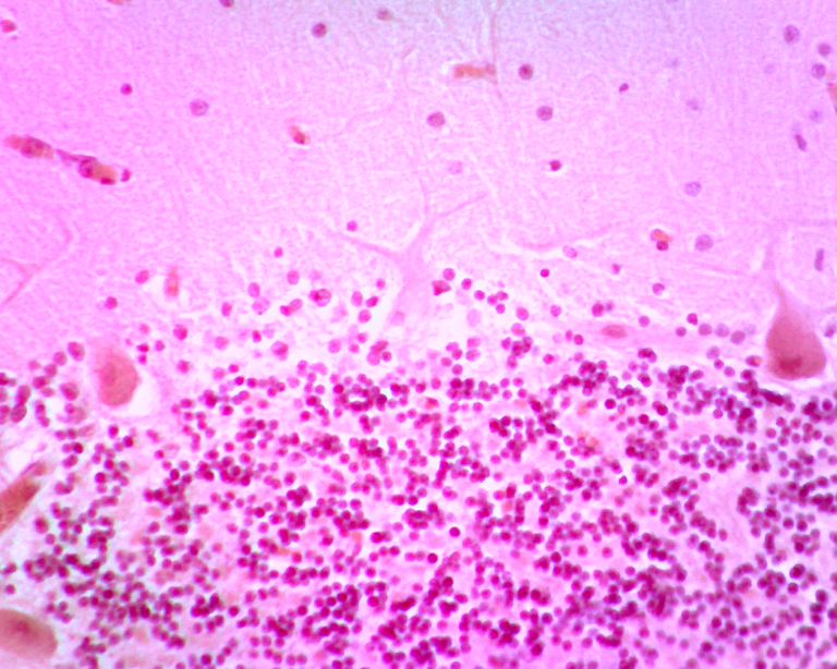
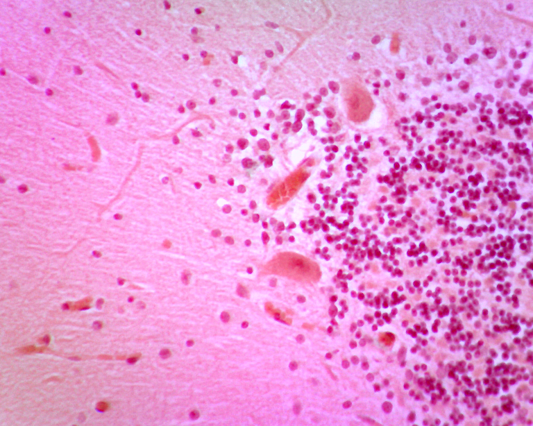
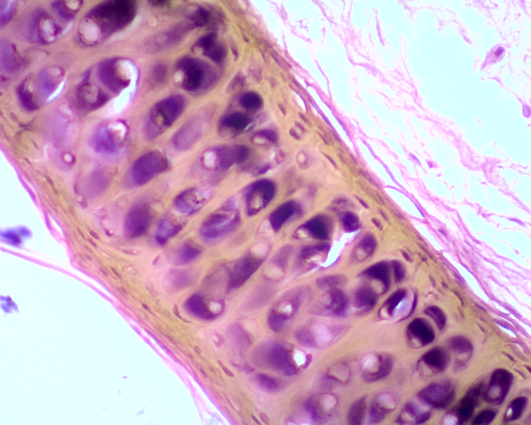
Nerve Tissue:
Nerve tissue is the tissue that makes up the central and peripheral nervous systems. This nerve cross section demonstrates how a tremendous concentration of nerve axons reside at the center of each nerve fascicle. A nerve fascicle is a bundle of faniculi, which are themselves bundles of nerve axons. The term nerve fascicle refers to nerves located in the peripheral nervous system; such structures located in the central nervous system are referred to as nerve tracts.
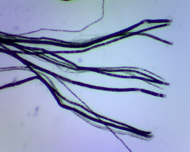
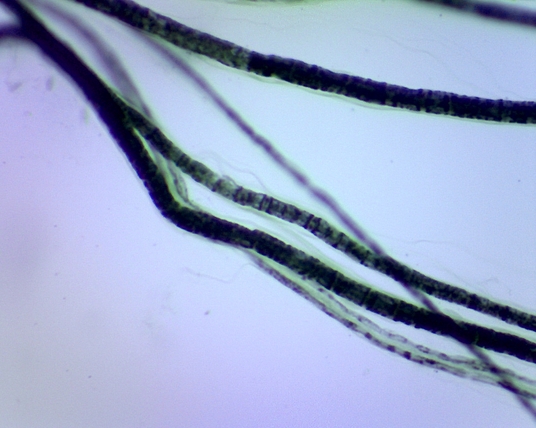
Nerve Fibers:
Nerve fibers are thin, elongated projections of nerve cells that are responsible for transmitting electrical impulses known as action potentials. Nerve fibers are subcategorized into Group A, Group B, and Group C nerve fibers based on their degree of myelination. Group A nerve fibers are heavily myelinated, Group B fibers are moderately myelinated, and Group C fibers are unmyelinated. Myelination refers to the presence of a myelin sheath surrounding a nerve cell’s axons. The myelin sheath in a lipid-rich (fatty) substance that insulates the axon to increase the speed of transmission of an action potential along the length of the axon. As a result, more heavily myelinated axons can transmit information more quickly that those with relatively less myelination.

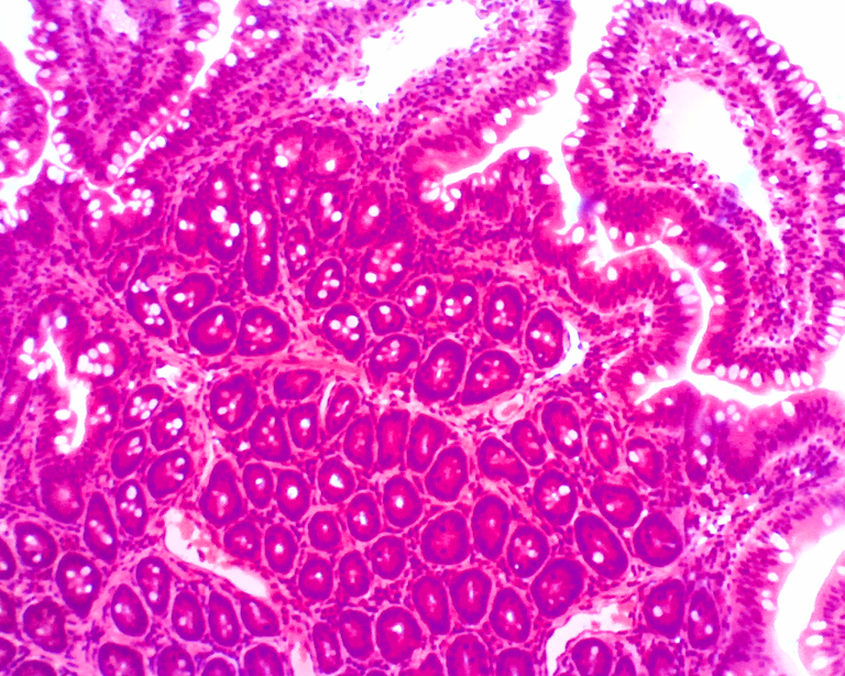
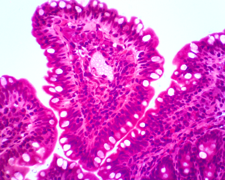
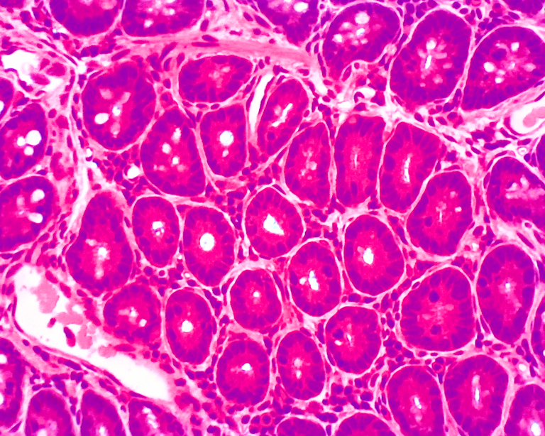
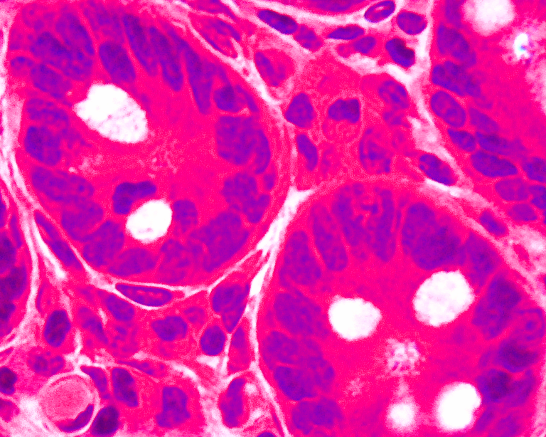
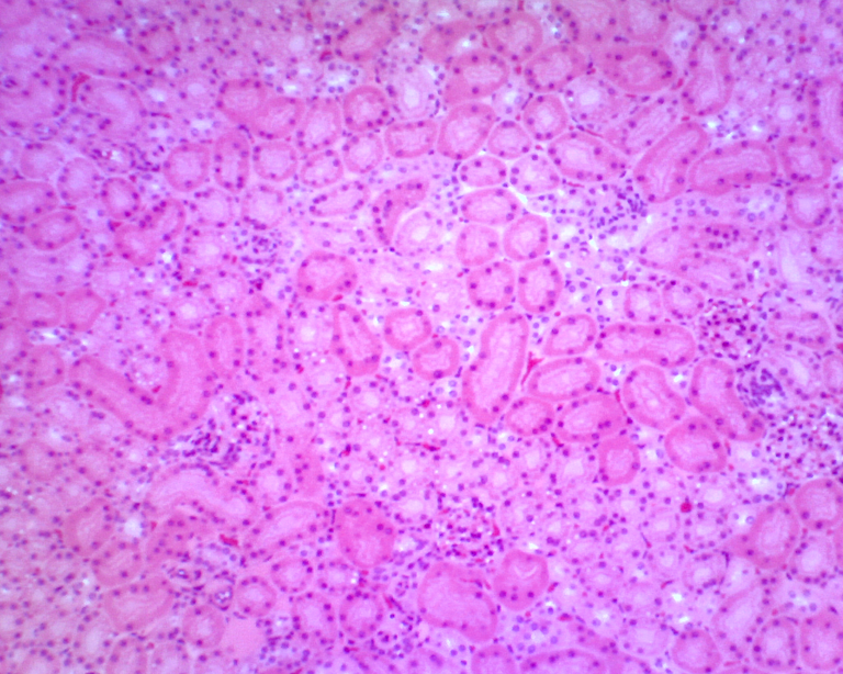
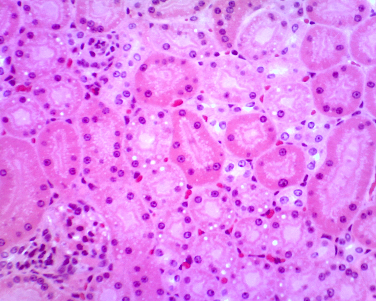
Simple Columnar Epithelium:
Simple columnar epithelium is a singular layer of tall, column-shaped cells that commonly line internal structures of the digestion system such as the stomach and intestines. The primary function of these cells in protective. For instance, the simple columnar epithelium that line the intestinal tract act as an impermeable barrier to bacteria while still allowing for the absorption of nutrients from food. Simple columnar epithelium are also responsible for secretion of acid, enzymes, and mucus in the stomach.
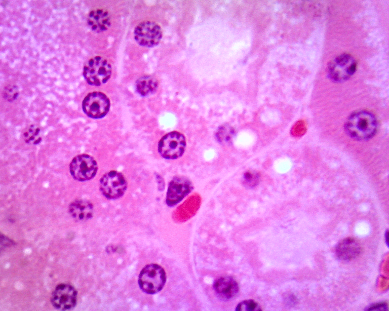
Simple Squamous Epithelium:
Simple squamous epithelium are flat, thin, scale-shaped cells that are commonly found lining internal structures such as blood vessels and alveoli in the lungs. Due to their thin profile, these cells are relatively more permeable in comparison to other epithelial cells, making them effective barrier tissues in structures that require the rapid diffusion of small molecules across membranes.
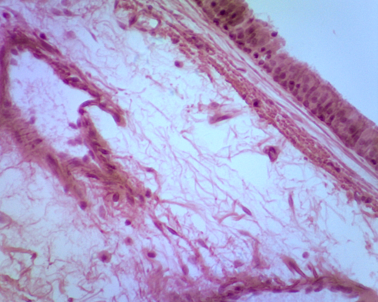
Trachea Cross-section:
The trachea, commonly referred to as the windpipe, is a cartilaginous tube that connects the larynx to the bronchi of the lungs. This rigid structure runs behind the breastbone and is responsible for conducting air from the nose and mouth into the lungs.

I hope that you enjoyed this microscopic view and that it gives you a better appreciation for the complex tissues that make up you and me alike!
My NFT Showroom gallery: https://nftshowroom.com/tych021/gallery
Creary Gallery: https://creary.net/@tych021/projects
Publish0x reflink: https://www.publish0x.com?a=M7e58kDYd2
PeakD reflink: https://peakd.com/register?ref=tych021
NFTShowroom reflink: https://nftshowroom.com/?r=tych021
Twitter: https://twitter.com/tych021
Vimm.tv: https://www.vimm.tv/tych021
Your content has been voted as a part of Encouragement program. Keep up the good work!
Use Ecency daily to boost your growth on platform!
Support Ecency
Vote for new Proposal
Delegate HP and earn more