Fungi Friday - Fungal Spore Photography
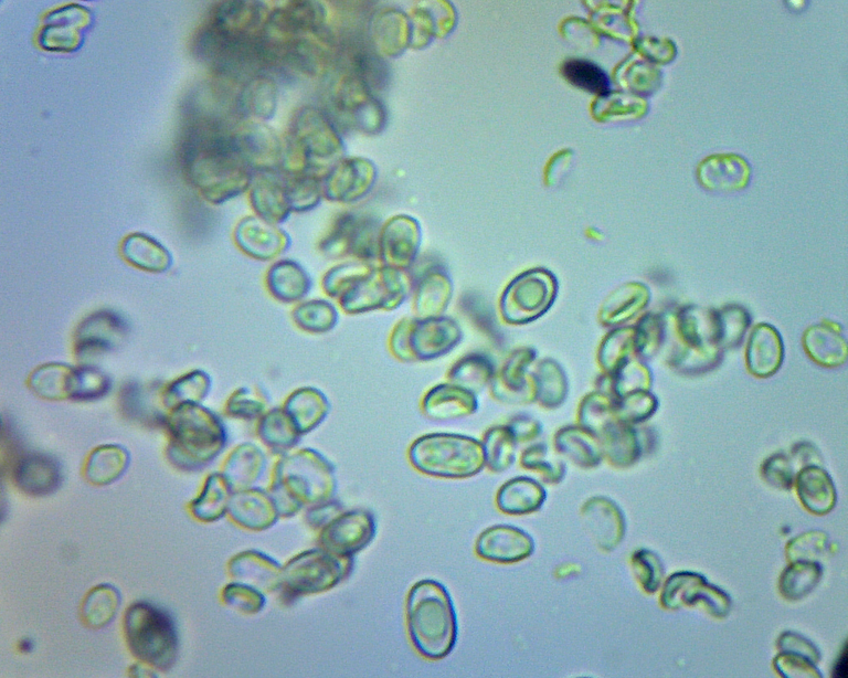
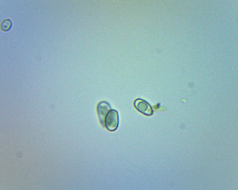
Happy Fungi Friday! Today I would like to share a few images taken of spores from a selection of fungi found in Western Michigan, USA. These images were taken over the course of the last few months as I continue to experiment with my microscopy setup.
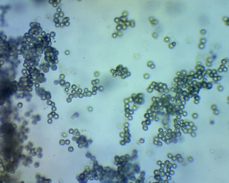
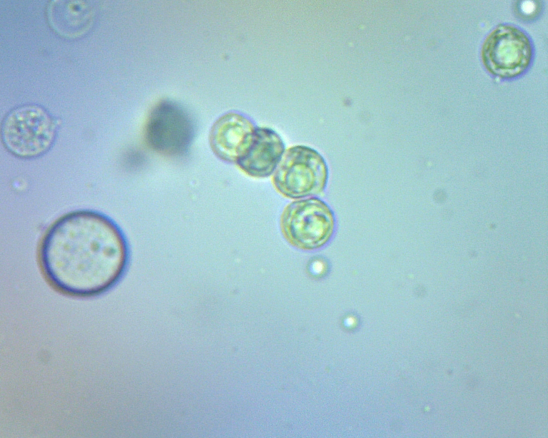
While my initial interest in fungi stemmed from the diversity of macrofungal forms, I am becoming increasingly interested in the fascinating variety of microscopic forms that can be found among fungal spores. I apologize for some of the blurry images shown below, but as I gain more experience with my setup, as well as upgrade my camera to a higher resolution, I am hoping to be able to document and share clearer images of spores collected from wild mushrooms.
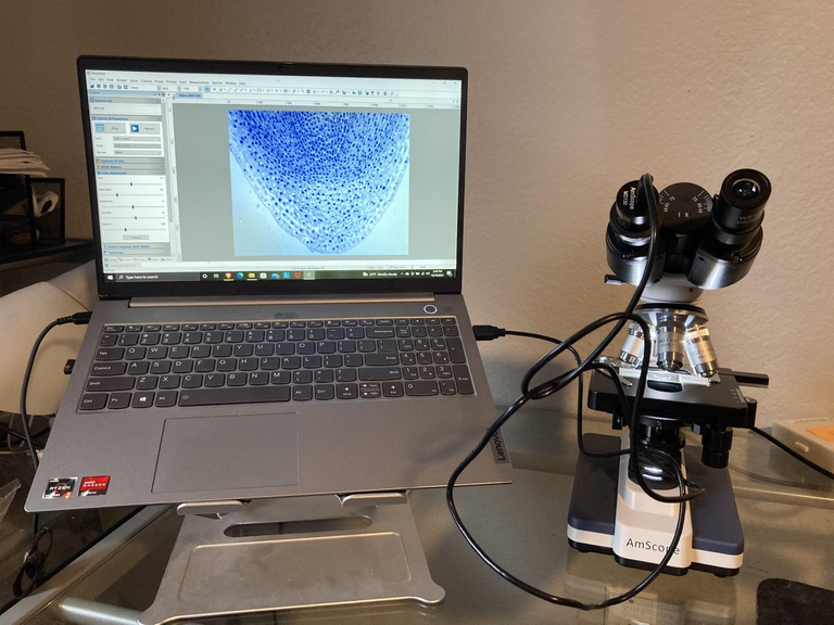
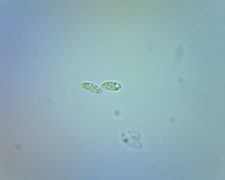
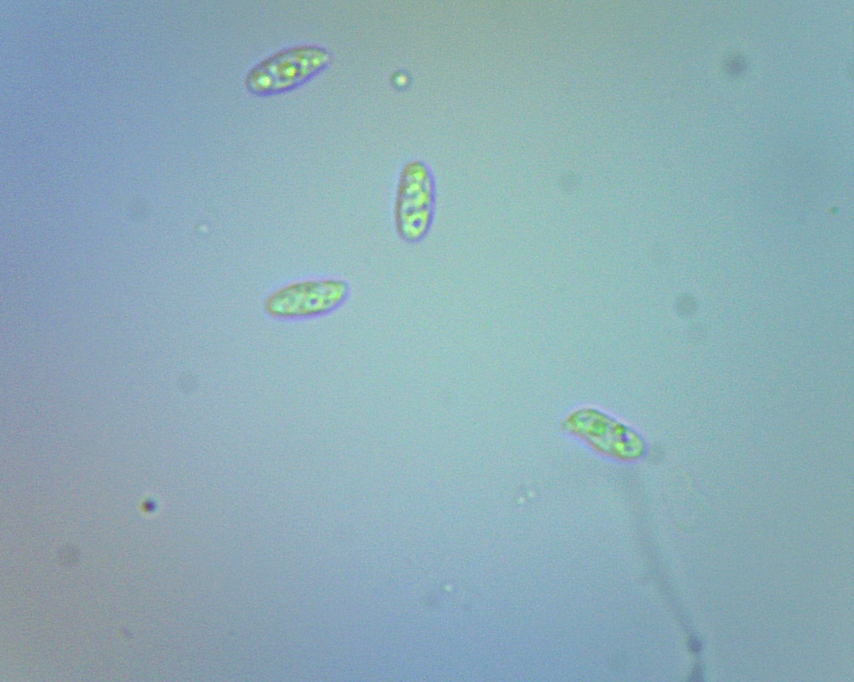
At the moment, I am wet-mounting spores collected from spore prints to view under an Amscope binocular compound microscope. This technique is great for viewing the spores in person, however, I have found that this is not ideal for capturing still images of the spores. Given the small size of the spores and the very narrow field of depth available at the level of magnification required to view these microscopic structures, it is often impossible to get an image that clearly displays the entirety of the spore in a single image. One work around that I have found for this issue is to take 10-20 images of the same spore while making miniscule adjustments to the fine focus knob on my microscope to adjust the field of depth through the entirety of the spore structure.
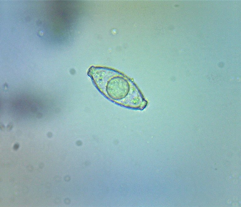
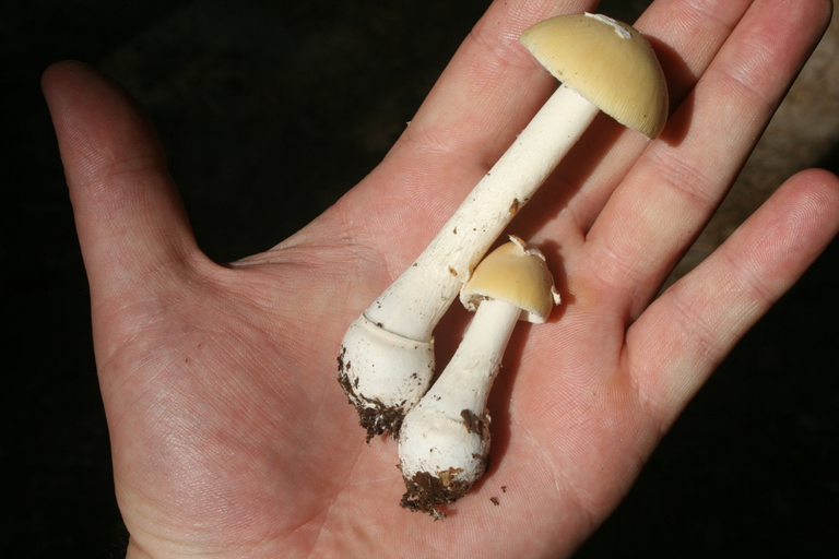
Early Spring Amanita (Amanita praecox) spore print:
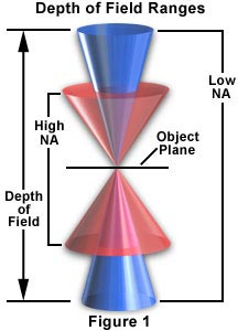
(Image source: https://micro.magnet.fsu.edu/primer/anatomy/focusdepth.html)
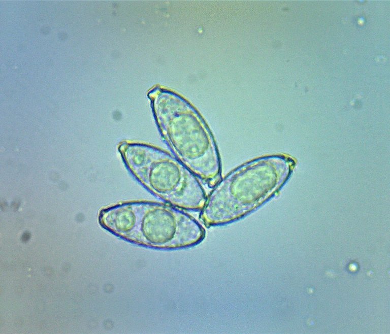
Once I have collected these images that focus on different depths within the spore stucutre, I can use an open-source software called CombineZP to combine the sharpest portions of each of the 10-20 images into a single, more crisp image of the spore. This is the technique employed to achieve the images shown for the Gyromitra Gigas complex spores. The downside of this technique is that it requires the spore to remain in one stationary position relative to the camera lens to allow for the images to properly stack. If the spores float around within the wet-mounted slide, they will not properly stack when run through CombineZP, and the resulting images will not provide a clear view of the spores. This is the precise problem I ran into while imaging the remaining spores shown in this post, which explains their relatively more blurred appearance in comparison to those of the Gyromitra gigas complex.
This all being said, my images are sufficient to show a small portion of the morphological diversity that exists among fungal spores. Some are globose, while others are more elliptical to cylindrical. Some possess germ pores and ornamentation, and others do not. While I am still learning the terminology used to describe the many forms that spores may take, and this post does not even begin to capture the vast diversity that exists among spore morphology, I am excited to discover new varieties and combinations as I delve deeper into this microscopic world. For any of those who may be interested in learning more about fungal spore morphology, I will provide a link here to a presentation deck created by Dr. Estelle Levetin of the University of Tulsa on Fungal Spore Morphology.
Happy Fungi Friday!
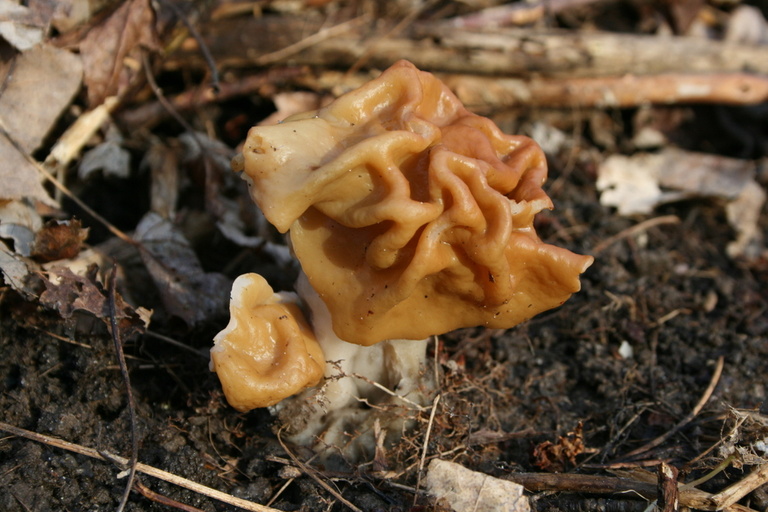
Gyromitra Gigas complex:
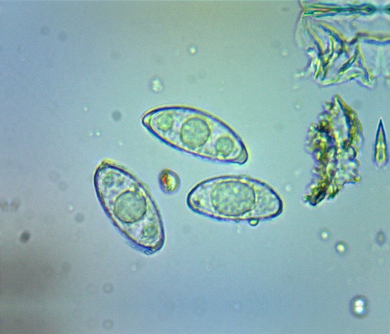
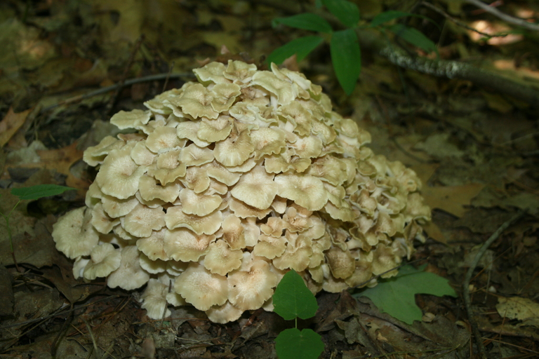
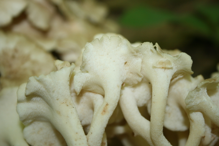
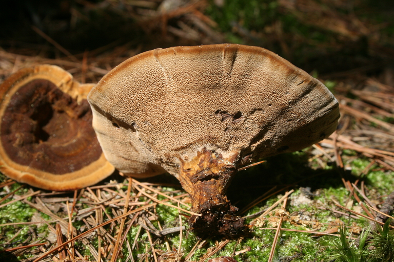
Umbrella Polypore (Polyporus umbellatus):
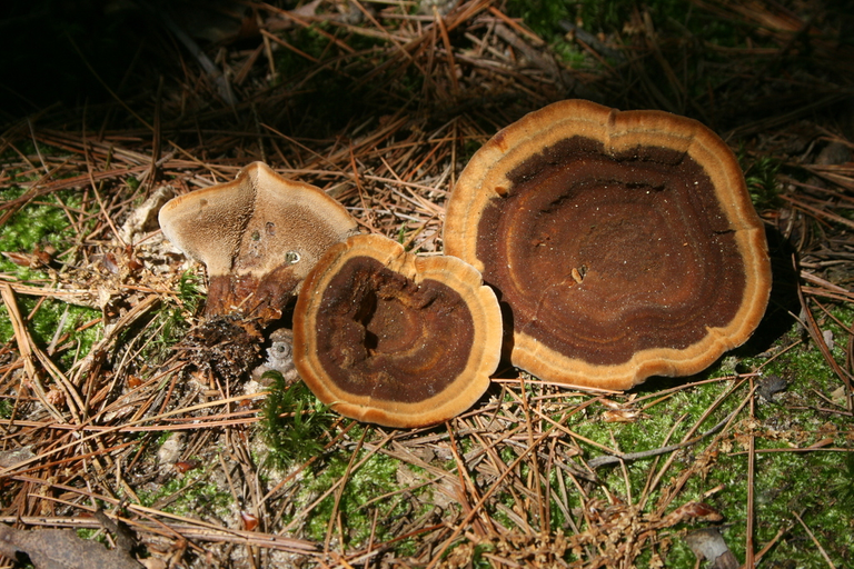
Dyer's Polypore (Phaeolus schweinitzii):
Early Spring Amanita (Amanita praecox):
My NFT Showroom gallery: https://nftshowroom.com/tych021/gallery
Creary Gallery: https://creary.net/@tych021/projects
Publish0x reflink: https://www.publish0x.com?a=M7e58kDYd2
PeakD reflink: https://peakd.com/register?ref=tych021
NFTShowroom reflink: https://nftshowroom.com/?r=tych021
Twitter: https://twitter.com/tych021
Vimm.tv: https://www.vimm.tv/tych021
it was a pleasure to walk with you, thanx for sharing quality stuff. the post is supported by FL upvote. if you like what community is about, please consider adding @hive-166168 as beneficiary of your posts -- lets grow together!
I totally forgot its Friday again!
Sneaks up on you sometimes!
It's a really great job you did on the mushrooms.
I also always miss the Friday, Thank you for sharing photos of how they look under microscope. Is it hard to make/spread mushrooms out in nature ? Like amadue
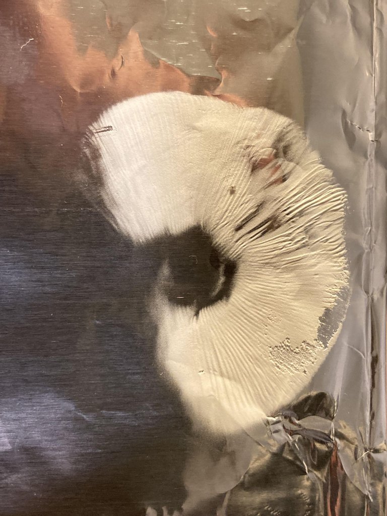
I'm not sure what you mean by making/spreading mushrooms our in nature, but if you are asking about collecting spores to grow mushrooms at home, it is fairly simple. You can collect spores from most cap and stem mushrooms by placing the cap on a piece of tin foil and placing a cup over the top to keep humidity up around the cap. Those spores can be used to make a spore syringe or grow the mycelium out on agar.
Thank you! Yeah I'm learning, but really inspired by the wonderful fungi's