The C.E.O of the Body - Cerebral Cortex 2: The Association Area's and lesion
In the previous blog, we looked at the projection areas of the cerebral cortex, and how they receive sensory Information and give out motor commands. But did you know that less than one-quarter of the human cerebral cortex is devoted to these projection zones?
Yes!
The remaining parts are what we call association areas. These areas are involved in our higher mental functions such as planning, remembering, perceiving, thinking and speech. They don't receive direct sensory input or produce motor output, but they play a role in integrating all those information together to accomplish higher processing.
For example:
They link sensory inputs from the projection areas to stored memories. So whenever we see an object, color or anything else, as it's received by the projection areas, it's linked to our previously stored memory in the association areas, which tells us what the object we are looking at is, as we've learned previously. This is generally what happens, although I must mention that there are more complex processes that takes place between sensory inputs and memory retrieval.
Now a lot of these information about how these processes work in the association areas of the cerebral cortex, was learned by studying people who have had damage, which we call lesions(when a tissue or organ suffer damage), from things like tumors, bleeding, accidents or blocked blood vessels (known as a stroke).
Additional evidence came from a comparison of the anatomy of the cortex found in different mammals. For example in the rats, a bigger region of the cortex is taken up by projection areas, while in cats, as we saw in the previous blog, proportionally more cortical space is devoted to the association areas. The proportion is even greater in monkeys, and when it comes to humans, it's greatest.
The usual understanding we had of what happens when there's damage in association areas is that it messes up how messages are organized from the sensory projections or to the motor projections. But we didn't know how to precisely figure out where these damages are? To be sure of their exact location, it would will eventually be through an autopsy.
But who would be the sacrificial lamb for this experiment?
Definitely not the patient, nor the scientist (he has to record the information and keep the study going). So both would refer to get an answer while they are still alive.
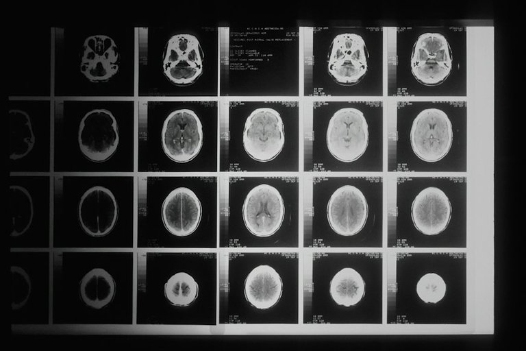
Then came standard X-rays which were of some help, but the problem was they only reveal major or obvious health issues. Luckily, we now have advanced methods that give us a more accurate view of the structure of a person's brain while they're alive.
One method is the CAT scan (Computerized Axial Tomography). It uses a thin X-ray beam directed through the head, hitting a detector on the other side. The beam circles around the head, and the detector moves too. Since brain tissues have different densities, they block X-rays in different ways. Finally, a computer puts together a complete picture using the X-ray views from all those different angles.
Then came even newer advancements, which enabled neurologists and psychologists to examine a live brain and observe certain aspects of how it works. The PET scan (Positron Emission Tomography) is one such technology, relying on the idea that active brain tissue, like any living tissue, consumes more energy.
What happens is, the person gets a shot of radioactive sugar that looks like glucose (the only fuel the brain uses), then the active brain cells take up this substance, emitting subatomic particles to show their presence. The PET scan then reveals the level of activity in different brain regions. High emission may indicate abnormal activity, suggesting issues like tumors, lesions, or psychological disorders.
Cool right?
Okay, so we've learned how modern neuropsychologists can figure out exactly where there's damage in the brain. Now, the question is:
What do these damages tell us about the jobs of the association areas in the brain's outer layer (cerebral cortex)?
There are certain damages(lesions) that can cause a situation known as apraxias. These are significant issues with starting or organizing voluntary actions.
In some cases of apraxias, the person is not able to do certain common actions like saluting or waving goodbye when requested. In other cases, actions that most people find simple and straightforward becomes a problem. A very popular example, is when a patient is asked to light a cigarette, the individual might repeatedly strike a match, even after it's already lit, or put the lit match into their mouth.
You see, these issues don't come from a lack of movement ability, as the person is well able to do each part of the action separately. The challenge lies in starting the sequence or choosing the correct components and putting them together.
Apraxia seems to be a problem with a neurological system that arranges individual movements into meaningful, bigger actions and starts them. Now, let's relate this to what we discussed in my blog two days ago about the hierarchical principle. We looked at transected cats and saw how various brain levels organize limb movements into acts and acts into straightforward goal-directed sequences. The same concept applies to actions in the cortex.
Lets give an analogy:
The association region whose lesion(damage) would lead to the condition called apraxia is perhaps similar to the command post of a military unit, that draws up the battle plan and orders an attack. If the command post falls and no other takes its place, there can be no organized attack, even though the individual soldiers are still able to fire their guns and throw their hand grenades.
I hope the above analogy helps you to understand this better?
At the moment, there's a lot of discussion about where exactly this neural command post is in the brain or if there's just one or maybe more than one.
Today's Bus Stops Here:
Thanks for joining me in today's blogisode! I hope you enjoyed learning about these topics as much as I did. As mentioned yesterday, I truly enjoy sharing this knowledge and want to ensure my readers do too. If you have any suggestions about this subject or my blogs, please feel free to drop them in the comment section. I'll see you tomorrow for a continuation of this beautiful journey. Stay safe.

Reference and links:
https://medlineplus.gov/ency/article/007472.htm
https://www.cell.com/trends/neurosciences/fulltext/0166-2236(82)90219-3#:~:text=The%20term%20'association%20cortex'%20refers,their%20afferent%20and%20efferent%20connections.
https://www.ncbi.nlm.nih.gov/books/NBK11109/
https://www.medicalnewstoday.com/articles/153201
https://www.google.com/amp/s/www.healthdirect.gov.au/amp/article/pet-scan
Congratulations @serenecounsel! You have completed the following achievement on the Hive blockchain And have been rewarded with New badge(s)
Your next target is to reach 1750 upvotes.
You can view your badges on your board and compare yourself to others in the Ranking
If you no longer want to receive notifications, reply to this comment with the word
STOPThanks for your contribution to the STEMsocial community. Feel free to join us on discord to get to know the rest of us!
Please consider delegating to the @stemsocial account (85% of the curation rewards are returned).
Thanks for including @stemsocial as a beneficiary, which gives you stronger support.
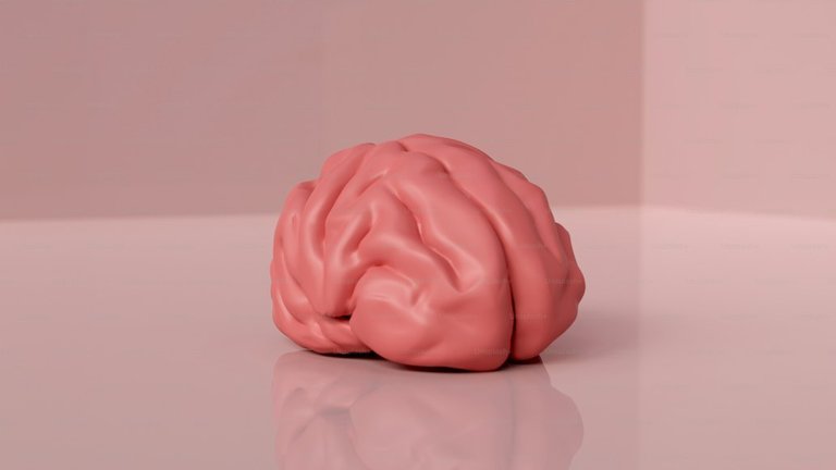
The brain, a powerful but delicate organ.
And there again we have the brains ability to rewire itself in the advent of any damage (neuroplasticity) through a neuroregenerative process