🦷The five X-rays that should be taken during endodontic treatment. | Las cinco radiografías que deben realizarse durante una endodoncia. 🦷
La endodoncia es conocida por todos como el tratamiento de conducto y es uno de los tratamientos endodonticos de mayor complejidad ya que hay que proteger no sólo el diente durante el procedimiento sino a cada uno de los tejidos que rodean al diente como lo es el periodonto, debemos cuidar nos pasarnos de la constricción apical que es ese espacio de cemento y dentina radicular, puesto que si con las limas perferoramos el paciente puede manifestar una sintomatologia que este no presentaba, más aún si posee un diagnóstico de necrosis pulpar ya que podemos arrastrar esas bacterias que están constituidas de material necrotico y originar abscesos, dolor e inflamaciones.
Endodontics is known by all as root canal treatment and is one of the most complex endodontic treatments because we must protect not only the tooth during the procedure but each of the tissues surrounding the tooth as is the periodontium, we must take care not to exceed the apical constriction that is the space of cement and root dentin, since if with the files we perferoramos the patient can manifest a symptomatology that this one did not present, even more if it has a diagnosis of pulp necrosis since we can drag those bacteria that are constituted of necrotic material and originate abscesses, pain and inflammations.
Es por ello que son muy importantes las radiografías, sino poseemos aparato de rayos X, no podemos hacer la endodoncia ya que estamos trabajando prácticamente a ciega, las radiografías nos permite evidenciar cada uno de los procedimientos y que lo estemos haciéndo de una manera correcta, por medio de esta publicación les hablaré de cuáles son esas radiografías que se realizan durante el tratamiento de conducto, las cuales son cinco.
That is why x-rays are very important, if we do not have an x-ray device, we cannot perform endodontics because we are working practically blind, x-rays allow us to show each of the procedures and that we are doing it in a correct way, through this publication I will talk about which are those x-rays that are performed during root canal treatment, which are five.
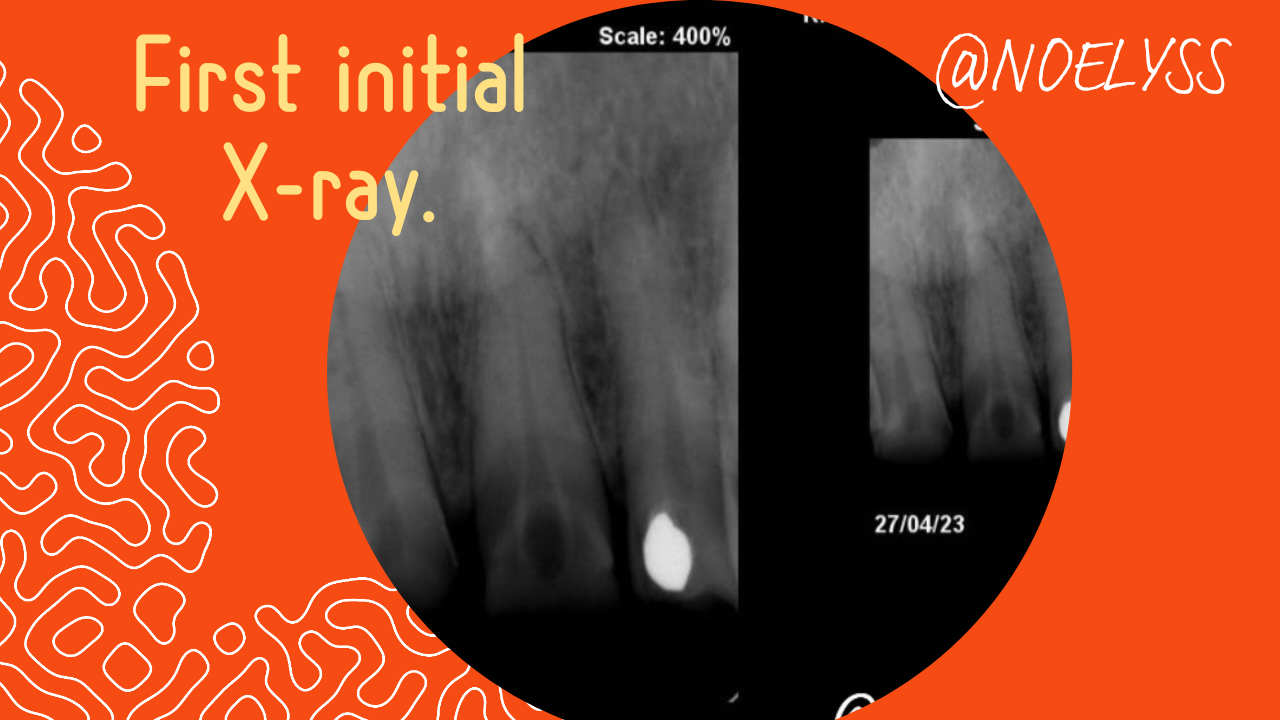
- Radiografía inicial: Esta radiografía debe realizarse antes del tratamiento de conducto, es un gran elemento diagnóstico ya que esta nos ayuda en el llenado de la historia clínica de endodoncia, permitiendo orientarnos en nuestro diagnóstico, su importancia radica en que ella nos permite tener una longitud tentativa que quiere decirnos cuanto mide aproximadamente a lo largo nuestro tratamiento de conducto, esta longitud tentativa debe ser colocada en la historia clínica mediante su medida, ya que nos ayudará para nuestra conductometria.
- Initial radiography: This radiography should be performed before the root canal treatment, it is a great diagnostic element since it helps us in the filling of the endodontic clinical history, allowing us to orient ourselves in our diagnosis, its importance lies in the fact that it allows us to have a tentative length that wants to tell us how long our root canal treatment is approximately, this tentative length should be placed in the clinical history by measuring it, since it will help us for our conductometry.
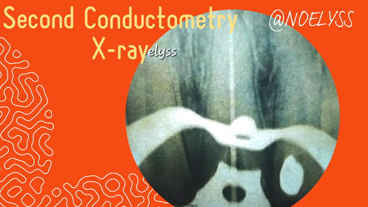
- Conductometría: Esta radiografía es una de las más importantes durante el tratamiento de conducto, porque nos permite determinar la longitud de trabajo, es decir la medida exacta de nuestro conducto que nos indica que instrumento debemos usar. Se determina de la siguiente forma, con una lima de 15mm y tomando en cuenta nuestra longitud tentativa, desde el borde incisal o la cupide más alta de un molar colocamos nuestro tope de goma como referencia en inducimos la lima dentro del conducto, y debemos verificar que la lima quede exactamente un milimetro por encima de la constricción apical en la radiografía, nuestra conductometria siempre debe ser exacta nunca debemos pasarnos, realizamos nuestro ajustes sumanos o restamos en caso de ser necesario hasta que tengas la medida exacta.
- Conductometry: This radiography is one of the most important during root canal treatment, because it allows us to determine the working length, i.e. the exact measurement of our canal, which tells us which instrument to use. It is determined in the following way, with a 15mm file and taking into account our tentative length, from the incisal edge or the highest cupid of a molar we place our rubber stopper as a reference and we induce the file inside the canal, and we must verify that the file is exactly one millimeter above the apical constriction in the radiograph, our conductometry must always be exact, we must never overdo it, we make our adjustments adding or subtracting if necessary until we have the exact measurement.
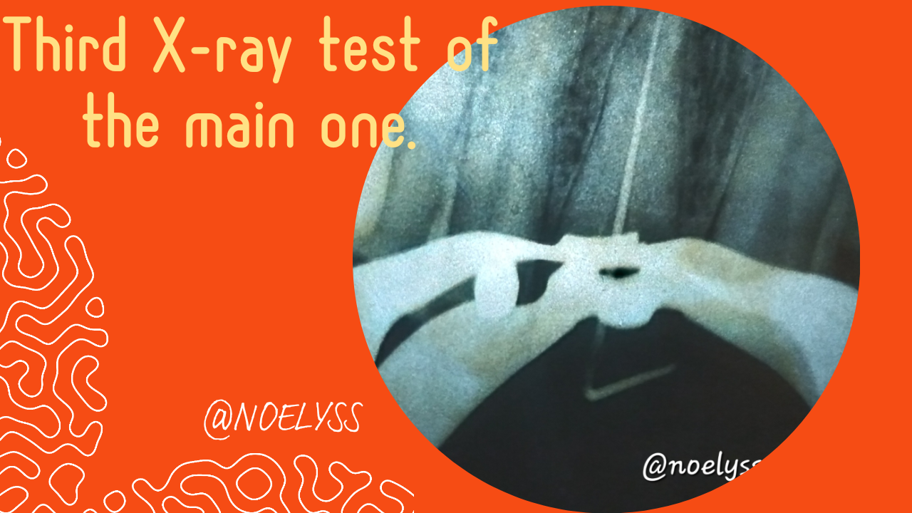
- Prueba del cono principal: Una vez que ya se haya preparado el conducto e instrumentando correctamente eliminando por completo el téjido pulpar infectado, procedemos a relizar nuestra prueba del cono principal esta se hace con una gutapercha del mismo tamaño de nuestra lima memoria, que fue lima con la cual preparamos hasta apical, mediante la prueba visual y táctil determinamos que nuestro cono de gutapercha principal este totalmente centrado a lo largo del conducto y que tenga un buen tamaño, debe ser del tamaño de nuestro conducto, no tan fino, se procede a tomar la radiografía.
- Main cone test: Once the canal has already been prepared and instrumented correctly eliminating completely the infected pulp tissue, we proceed to perform our main cone test this is done with a gutta percha of the same size of our memory file, which was file with which we prepared up to apical, by visual and tactile test we determine that our main gutta percha cone is totally centered along the canal and that it has a good size, it must be the size of our canal, not so thin, we proceed to take the radiograph.
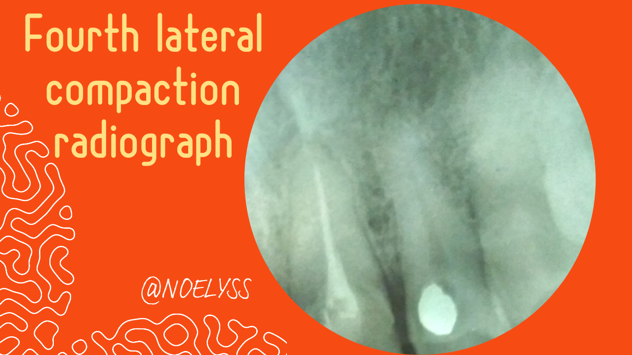
- Compactacion lateral: Después de tomar la radiografía de cono principal seguimos con la compactacion una vez uue tenemos seleccionado nuestro cono principal, colocamos nuestras gutaperchas accesorias y compactamos muy bien cada una, permitiendo que el el cemento obturador pueda adherirse bien a la paredes logrando un sellando hermético, justamente al terminar de compactar se toma la radiografía y nos garantizamos que no se encuentren espacios radiolucidos lo que puede indicarnos que debemos agregar más gutaperchas, es muy necesario que el conducto quede bien obturado, sin espacios o filtración, esta radiografía no es más que el control de la obturación del conducto radicular.
- Lateral compaction: After taking the main cone x-ray we continue with the compaction once we have selected our main cone, we place our accessory gutta percha and compact each one very well, allowing the obturator cement to adhere well to the walls achieving a hermetic seal, Just when we finish compacting, we take the x-ray and we make sure that there are no radiolucent spaces which may indicate that we should add more gutta percha, it is very necessary that the canal is well sealed, without spaces or filtration, this x-ray is nothing more than the control of the obturation of the root canal.
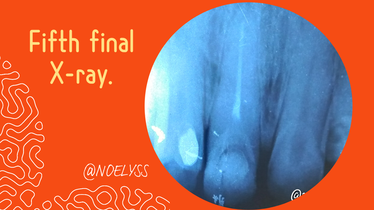
- Radiografía final: Una vez que ya se hayan realizado cada unos de los pasos o procedimientos del tratamiento de conducto, vamos a realizar nuestra relación final que la misma debe quedar bien hecha, es de gran importancia ya que la misma nos indica el éxito del tratamiento endodontico.
- Final X-ray: Once each of the steps or procedures of the root canal treatment have been carried out, we are going to make our final report, which must be well done, it is of great importance since it indicates the success of the endodontic treatment.
Por último es muy importante que tengamos en cuenta como pacientes estas cinco radiografías que se deben realizar durante un tratamiento de conducto, ya que existen odontólogos que no siguen este protocolo por lo que existen mas probabilidades el fracaso endodontico del tratamiento, espero esta publicación sea de tu interes gracias por leerme
Finally, it is very important that we as patients take into account these five x-rays that should be taken during a root canal treatment, since there are dentists who do not follow this protocol, so there are more probabilities of endodontic treatment failure, I hope this publication is of your interest, thanks for reading me.
Referencia Informativa/Reference Informative:
https://estudidentalbarcelona.com/radiografia-en-endodoncia-cual-es-la-ideal/

Texto traducido en Deelp
It's very important to have one. The machine look very great to execute any hidden problem especially when it comes to internal structure.
Thanks for your contribution to the STEMsocial community. Feel free to join us on discord to get to know the rest of us!
Please consider delegating to the @stemsocial account (85% of the curation rewards are returned).
You may also include @stemsocial as a beneficiary of the rewards of this post to get a stronger support.