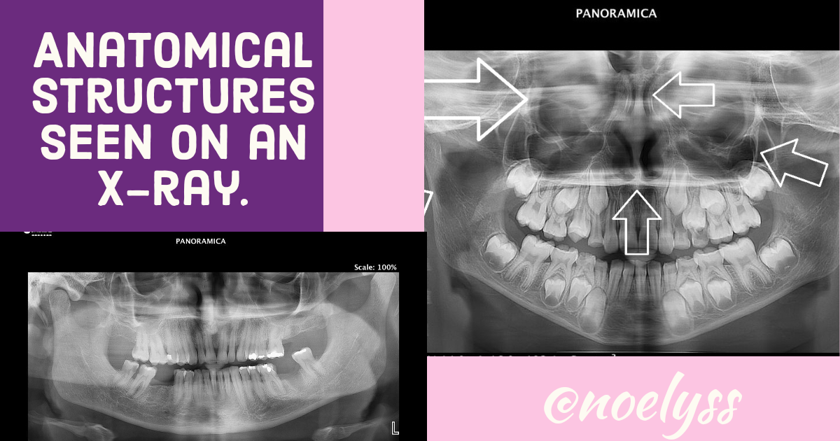
💀Anatomical structures seen on an X-ray | Estructuras anatomicas que observan en una radiografía panorámica.💀
My friends of this community I send you all a big hug, for some weeks I have not published in this community I have been a little discouraged but today I bring you my content of dentistry, panoramic radiographs are essential for the diagnosis in dentistry as they allow us to show what we can not observe clinically, so it is necessary that for any dental treatment the patient can take his panoramic radiography, This radiography allows us to observe various anatomical structures not only the hard and soft tissues of the oral cavity but also nasal anatomical structures that in some cases allows us to realize that the patient about sinositis among others, so in this publication I will talk about the anatomical structures that can be observed in the same.

Para identificar las estructuras anatomicas de la radiografía panorámica primeramente debemos hacerlo en el maxilar superior porque posee mayor detalles anatómicos que en el maxilar inferior, una vez que describimos e interpremos, lo hacemos en la mandibula.
To identify the anatomical structures of the panoramic radiograph we must first do it in the upper jaw because it has more anatomical details than in the lower jaw, once we describe and interpret, we do it in the mandible.
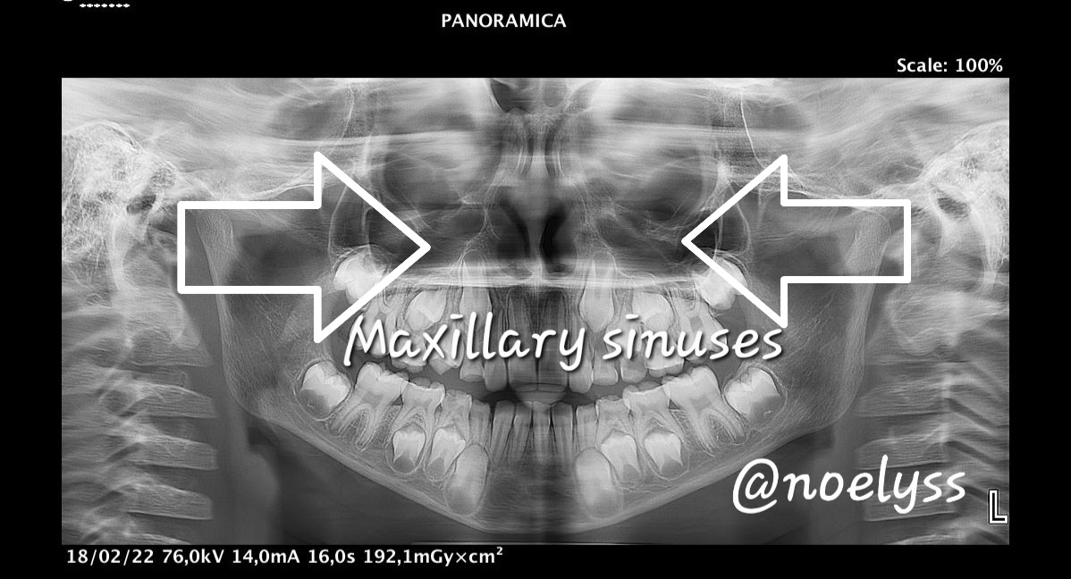
El hueso maxilar superior, es esponjoso trabeculado, este tiene varios detalles anatomicos importantes en la radiografía panorámica como lo es el seno maxilar, que es una cavidad neumática que contiene aire, su principal función es la misma, es muy importante poder observarlo ya que debido a caidas o diversos traumatimos el paciente puede tener el mismo totalmente descendido, por lo que conlleva que las raíces de los molares puedan estar dentro del seno y se debe complentamente hacer un correcto abordaje quirúrgico puesto que puede haber una comunicación bucosinosal en el paciente y el mismo puede quedar, arrojando sangre debido a la comunicación que posen esas estructuras con las fosas nasales.
The upper maxillary bone is trabeculated spongy, it has several important anatomical details in the panoramic radiography such as the maxillary sinus, which is a pneumatic cavity containing air, its main function is the same, it is very important to observe it because due to falls or various traumas the patient may have it completely descended, Therefore, the roots of the molars can be inside the sinus and a correct surgical approach must be made, since there can be a buccosinusal communication in the patient and the patient can be left, throwing blood due to the communication that these structures have with the nostrils.
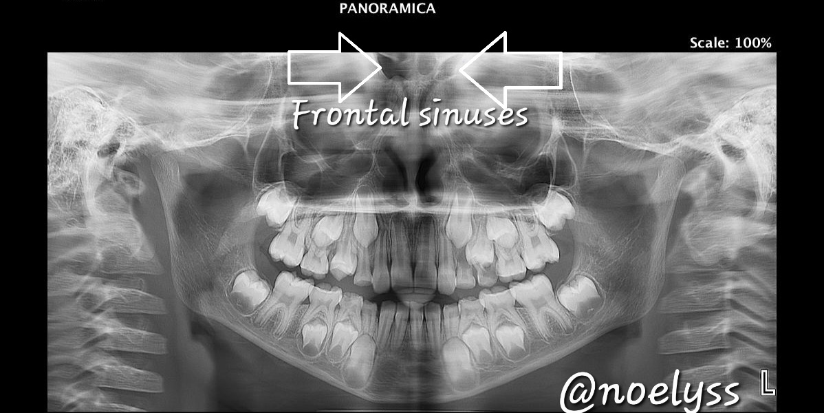
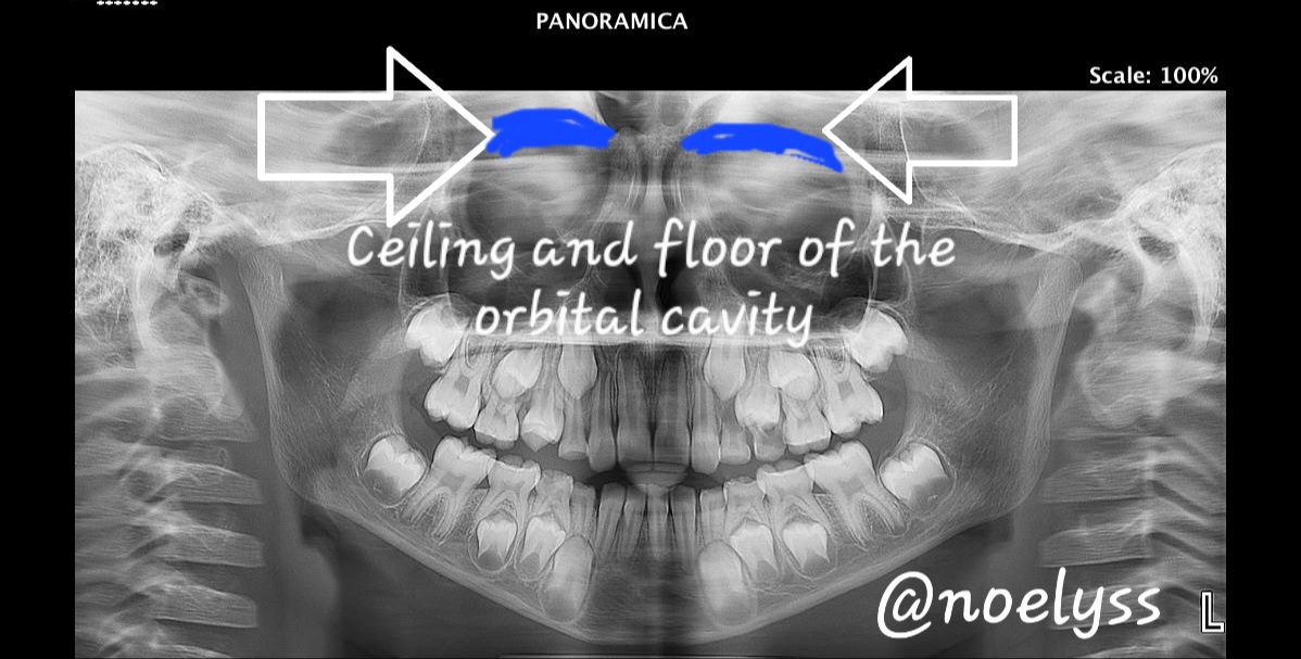
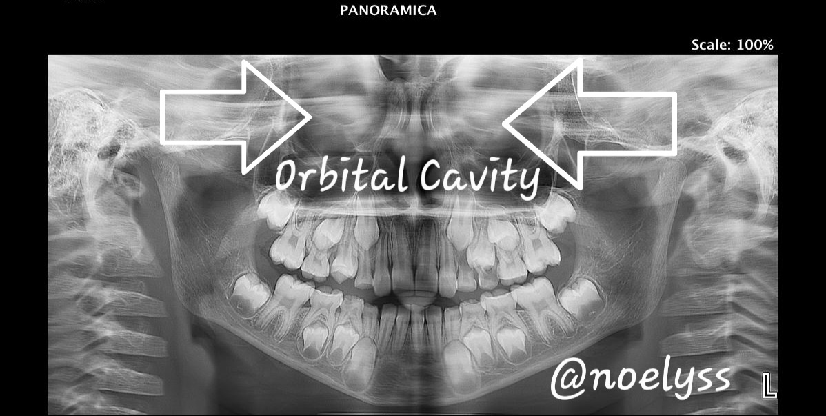
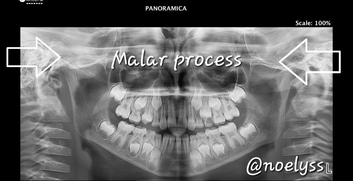
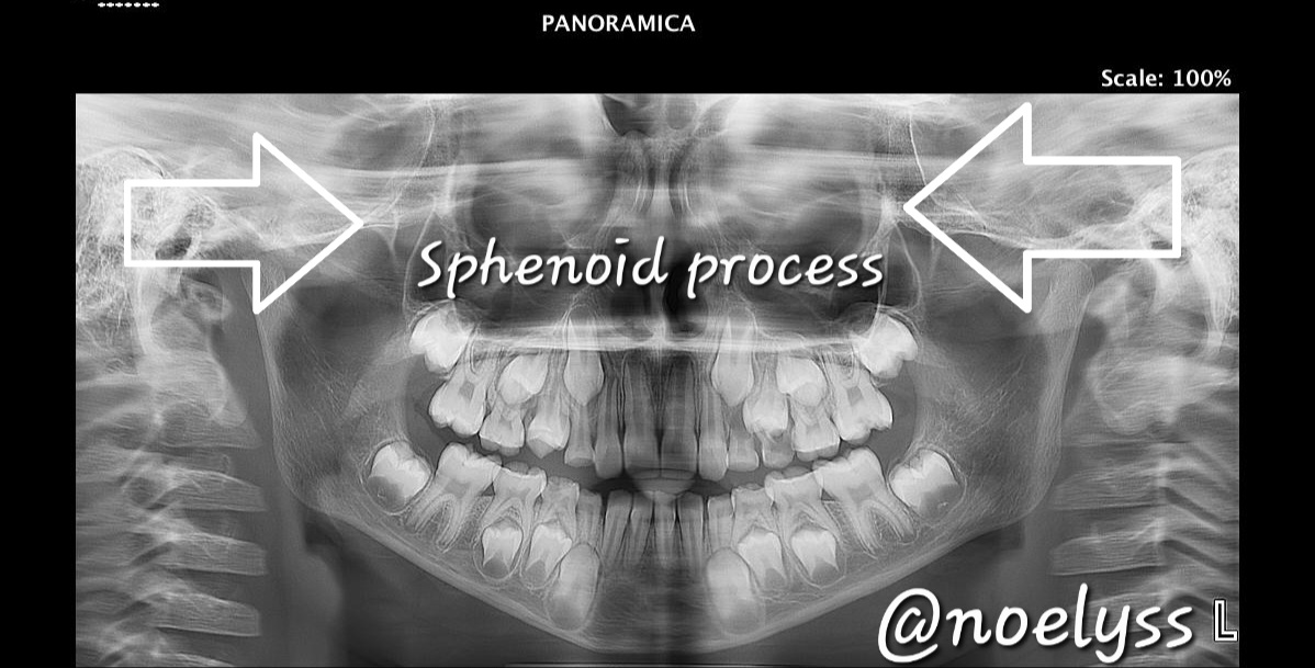
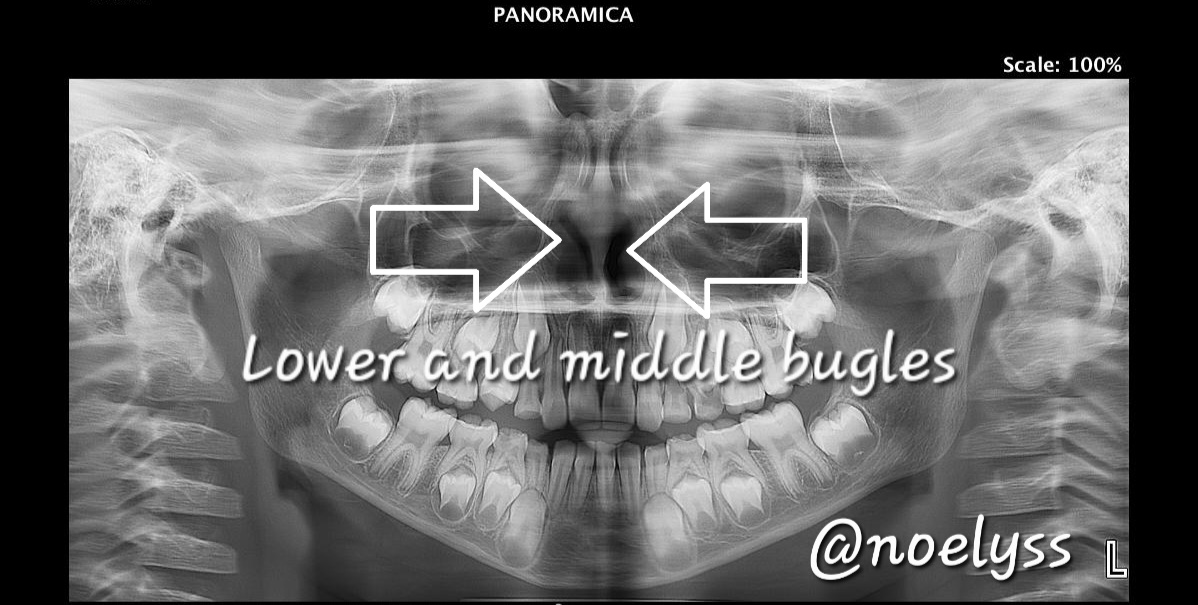
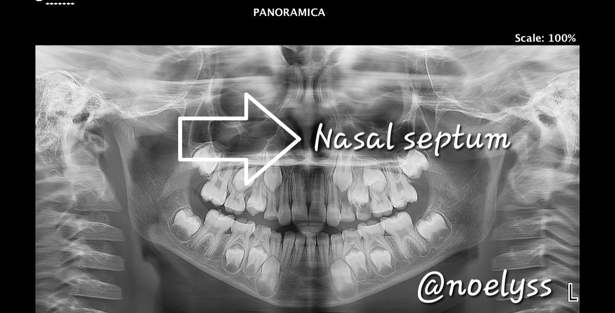
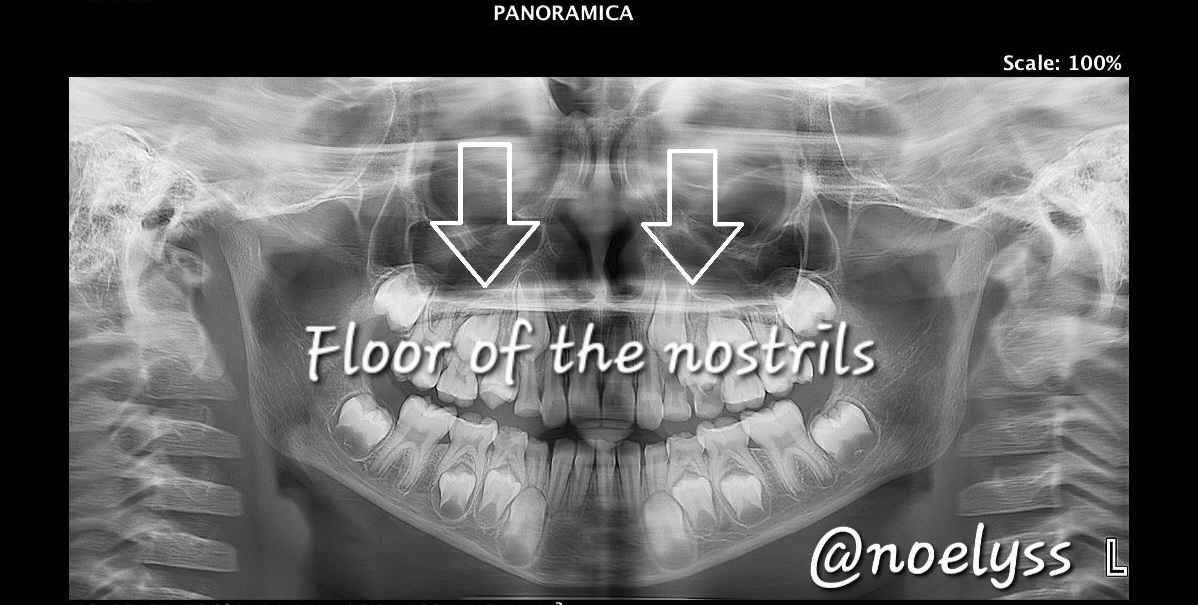
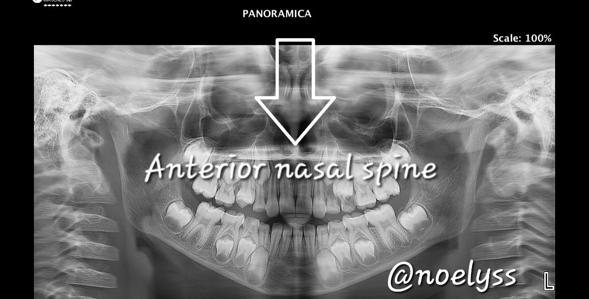
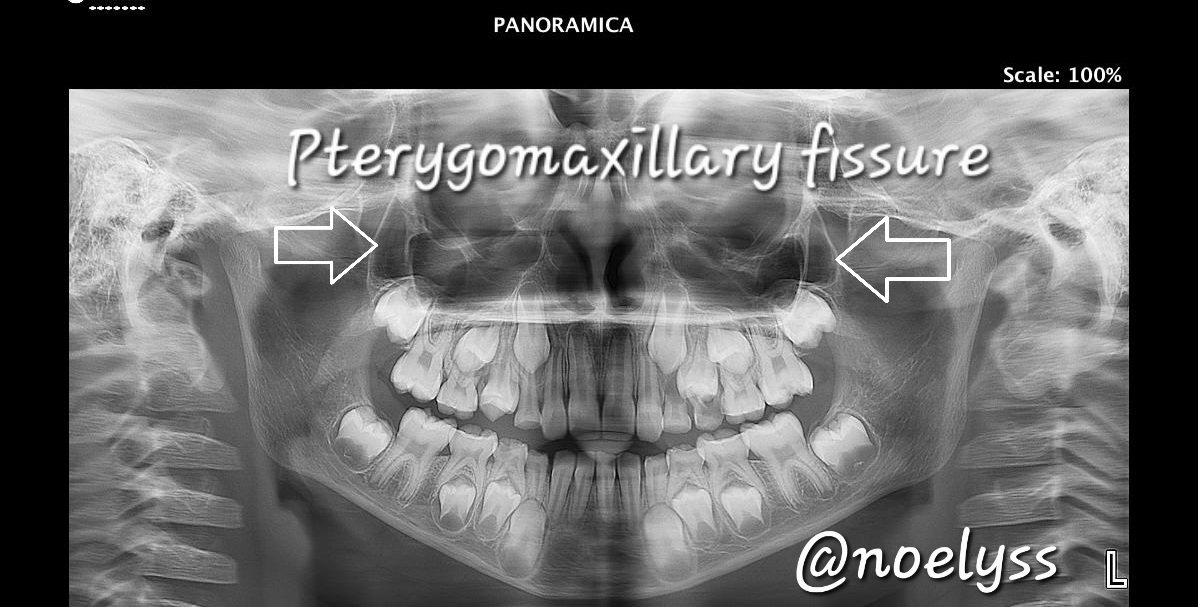
En las siguientes radiografías panorámicas podemos evidenciar una a una, todas las estructuras anatómicas que se conforman en el maxilar superior y otras muy importantes iniciando prineramente con los senos frontales que estos forman parte del hueso etmoides importantes en las filtración del aire, luego el piso y techo de las cavidades orbitarias que representa al hueso frontal craneo, y el piso al hueso malar, las orbitas muy importantes ya que se albergan los organos de los ojos en dicha cavidades, el septum nasal, la espina nasal anterior, los cornetes medios son involucrados y responsables en la respiración, en la radiografía los podemos observar cuando estos se encuentran de diferentes tamaño y así se puede dificultar un poco la respiración o en paciente, ya sea porque es alérgico.
In the following panoramic radiographs we can see one by one, all the anatomical structures that are formed in the upper jaw and other very important starting first with the frontal sinuses that are part of the ethmoid bone important in air filtration, then the floor and ceiling of the orbital cavities representing the frontal bone cranium, and the floor of the malar bone, the orbits are very important because they house the organs of the eyes in these cavities, the nasal septum, the anterior nasal spine, the middle turbinates are involved and responsible for breathing, in the X-ray we can see when these are of different sizes and thus can hinder breathing a little or patient, either because it is allergic.
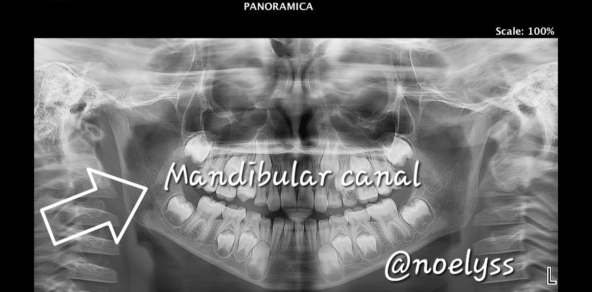
Ahora procedemos a conocer las estructuras anatomicas del hueso maxilar inferior. Es un hueso compacto de estructura mucho mas dura que de hecho podemos observar esta gran característica en la radiografía panorámica, cabe destacar que tiene detalles anatómicos muy importantes como el canal mandibular que va albergar el paquete vasculonervioso del nervio dental inferior, que el mismo permite la inervacion de media hemiarcada.
Now we proceed to know the anatomical structures of the lower maxillary bone. It is a compact bone of much harder structure that in fact we can observe this great feature in the panoramic radiograph, it is noteworthy that it has very important anatomical details such as the mandibular canal that will house the vasculonervous bundle of the lower dental nerve, which itself allows the innervation of half hemiarch.
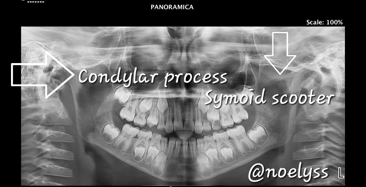
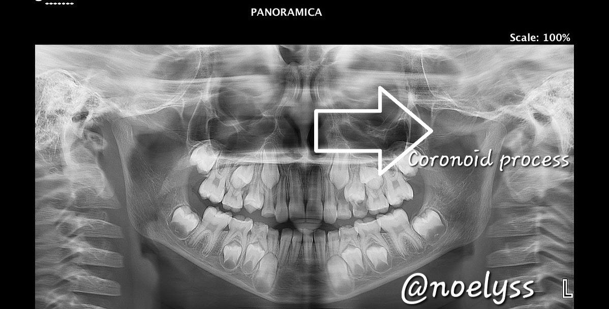
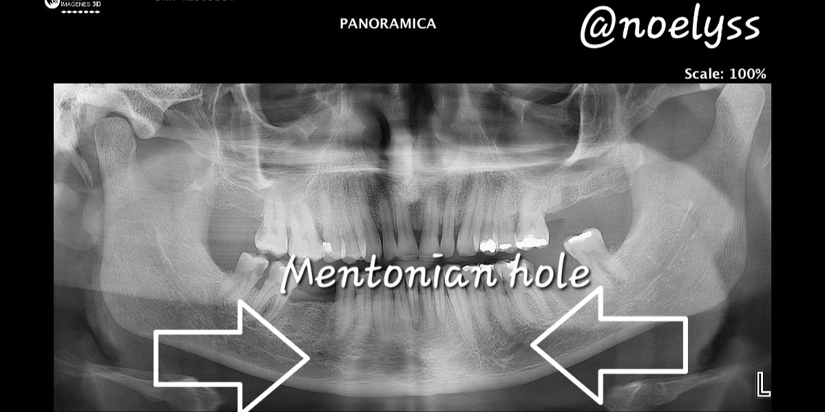
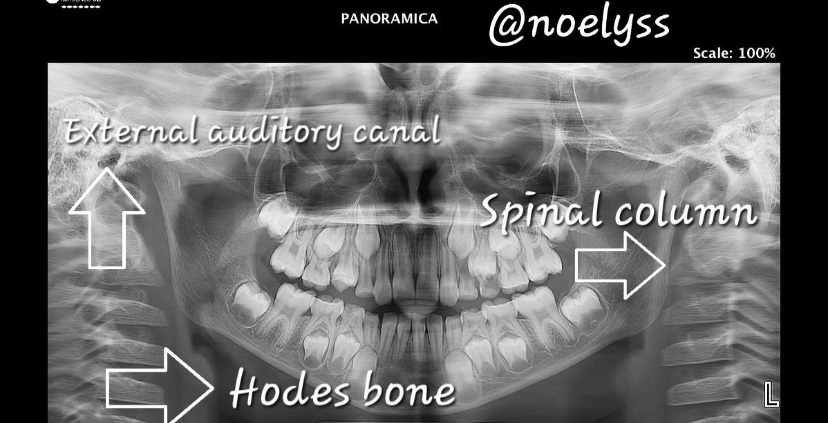
Podremos encontrar el condilo mandibular, que este permitira articular con la cavidad glenoidea del hueso temporar es redondo y se puede evidenciar perfectamente en la radiografía, así como también la escotadura simoidea que separa el condilo de la mandibula del proceso coronoide, los agujeros mentionanos que van a permitir el paso del nervio mentoniano, así como el conducto auditivo interno el orificio de entrada del oído, la columna vertebral se observa de perfecta manera a cada lado de la radiografía y por ultimo el hueso hiodes.
We can find the mandibular condyle, which will allow articulation with the glenoid cavity of the temporal bone is round and can be seen perfectly on the radiograph, as well as the symoid notch that separates the condyle of the mandible of the coronoid process, the mentionanos holes that will allow the passage of the mentonian nerve, as well as the internal auditory canal, the entrance hole of the ear, the spine is observed perfectly on each side of the radiograph and finally the hyoid bone.
Muchas gracias amigos queridos de esta comunidad de ciencias, espero hayan podido aprender las diversas estructuras anatomicas que se encuentran las radiografía panorámica, les envio un abrazo nos vemos en la próxima oportunidad.
Thank you very much dear friends of this science community, I hope you have been able to learn the various anatomical structures found in panoramic radiography, I send you a hug, see you next time.
Reference Informative:
(https://campusodontologico.com/radiografias-panoramicas/)
Texto traducido en Deelp
Thanks for your contribution to the STEMsocial community. Feel free to join us on discord to get to know the rest of us!
Please consider delegating to the @stemsocial account (85% of the curation rewards are returned).
You may also include @stemsocial as a beneficiary of the rewards of this post to get a stronger support.