THE FUNGUS: PNEUMOCYSTIS JIROVECII
During one of my Clinical posting I discovered an interesting fungus Formerly called, Pneumocystic carnii, the organism normally causes Pnemonia in immunocompromised human known as Pnemocystis Pnemonia (PCP). PNEUMOCYSTIS JIROVECII affect human as an opportunistic fungus that infect immunocompromised host, and mostly manifest as PCP. PCP is the most common infection in HIV patients . Pneumocystics is a unicellular fungus found in the respiratory tracts of many mammals and humans. The Genus Pneumocystics was initially mistaken a trypanosome, then was assumed to be a protozoan. Now later, biomedical analysis like nucleic acid composition of Pnemocystics ribosomal RNA and mitochondrial DNA established this Organism as definitely a fungus. The cyst wall closely resembles that of a fungus, however, it does not have a ergosterol in its membrane as typical as other fungi but instead has cholesterol.

Source:https://www.freepik.com/free-vector/mold-spores-grow-human-lungs_30551671.htm#fromView=search&page=1&position=1&uuid=4ab63871-b926-42ca-9eb0-35e024158fd5
Morphology
The Organism have three distinct morphological stages
- TROPHOZOITES: They are thin walled 1-5microlitre in size and is irregularly shape (amoeboid). It is thought to reproduce by binary fission.
- PRE CYST STAGE: The precyst is recognized as intermediate stage of the sexual phase of reproduction leading to cyst development. It is about 5-8microlire in size.
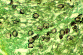
- CYST STAGE: they are thick walled and spherical about 8microlitre, contain up to eight (8) intracystic bldies.
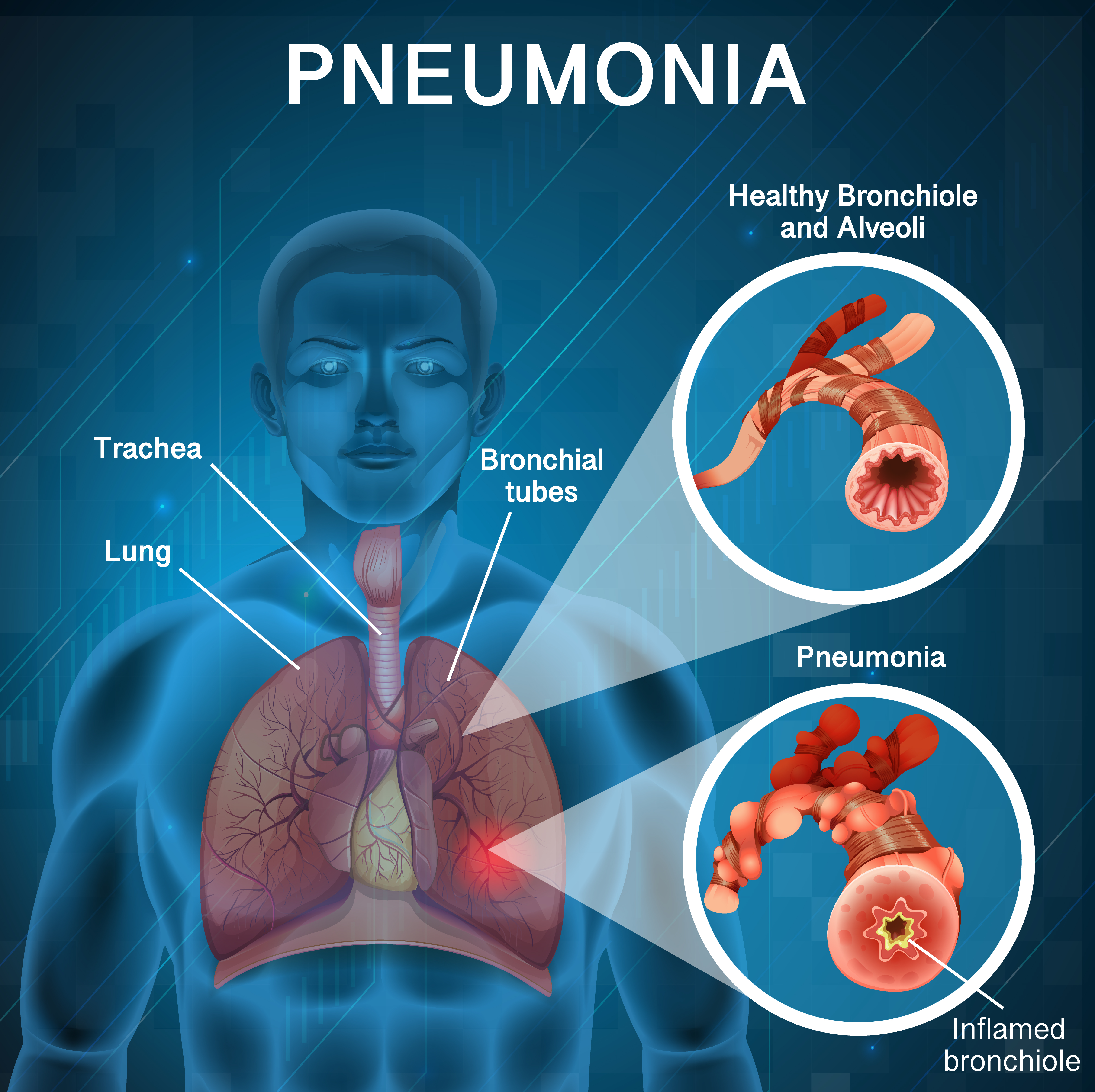
then lets look on how their work

Source: https://www.freepik.com/free-vector/poster-design-pneumonia-with-human-bad-lungs_7541561.htm#fromView=search&page=1&position=12&uuid=4ab63871-b926-42ca-9eb0-35e024158fd5

Source:https://www.freepik.com/free-vector/coronavirus-concept_7311140.htm#fromView=search&page=1&position=49&uuid=4ab63871-b926-42ca-9eb0-35e024158fd5
Pathogenesis
After Pnemocystic jirovecii is inhaled, the tropic form ( trophozoids) of the pathogen is believed to be adhered to type 1 Pnemocytes (thin squamous epithelial cells) of the lungs. The organism replicates extracellularly while bathed in alveoli lining fluids. With successful replication of the organism, the alveoli spaces filled with an eosinophilic foamy material which can be detected with H and E staing


