Simplifying the anatomy of the Upper limb: The proximal radioulnar joint
Introduction
Imagine a little girl waving her hands excitedly as a passenger airplane prepares for takeoff. In that moment of pure wonder and innocence, she marvels at the mechanics of her own hand, effortlessly creating the gestures that convey her excitement. Just like the coordination of her fingers and wrist in waving, the human body is a masterpiece of precision and complexity, especially in its joints. One such marvel is the proximal radioulnar joint, a component of the human arm that enables movements for daily activities. In this article, we will delve into the anatomy of the proximal radioulnar joint, discussing its structure and function.
Definition and the Importance
The proximal radioulnar joint is where the radius and ulna bones of the forearm meet near the elbow. This junction allows for the rotation of the forearm, which is important for everyday activities requiring movements like twisting, turning, and flipping. Think about how you use your forearm to turn a doorknob, stir a pot on the stove, or even just wave hello – all made possible by the flexibility and stability provided by the proximal radioulnar joint.
Without this joint, simple tasks like pouring a cup of coffee, typing on a keyboard, or even giving a thumbs-up would be much more challenging. The radioulnar joint's ability to rotate allows for a wide range of motion in the forearm, making it a key player in our daily routines. So, whenever you turn a key in a lock or give a friendly wave, remember to thank your proximal radioulnar joint for its hard work behind the scenes. Smile 😊
Articulation of the Proximal Radioulnar Joint
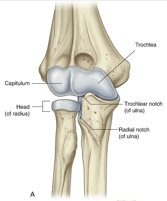
The proximal radioulnar joint is a pivot joint, which is a type of synovial joint that allows uniaxial rotation around a central axis. In this case, the radius bone rotates around its own axis, crossing over the ulna bone to enable pronation and supination of the forearm. This pivotal movement is controlled by the annular ligament, a strong band of fibrous tissue that encircles the radial head and holds it in place, facilitating smooth rotation without dislocation.
At the proximal radioulnar joint, the head of the radius bone articulates with the radial notch of the ulna bone, forming a stable but flexible connection. The articulation between these two bones is supported by ligaments and muscles that work synergistically to maintain proper alignment and function of the joint. The radial notch of the ulna acts as a socket for the radial head, allowing for smooth rotation without compromising stability.
The unique structure and function of the proximal radioulnar joint sheds light on its importance in our daily activities and highlights the design of our musculoskeletal system.
Capsule of the Joint
The capsule of the proximal radioulnar joint is a crucial structure that surrounds and supports the joint, providing stability while allowing for necessary movements like pronation and supination. This fibrous capsule is lined with synovial membrane, which secretes synovial fluid to lubricate the joint and reduce friction during motion. The capsule is reinforced by ligaments that help to maintain the integrity of the joint and prevent excessive movement that could lead to injury.
Ligaments surrounding the Joint
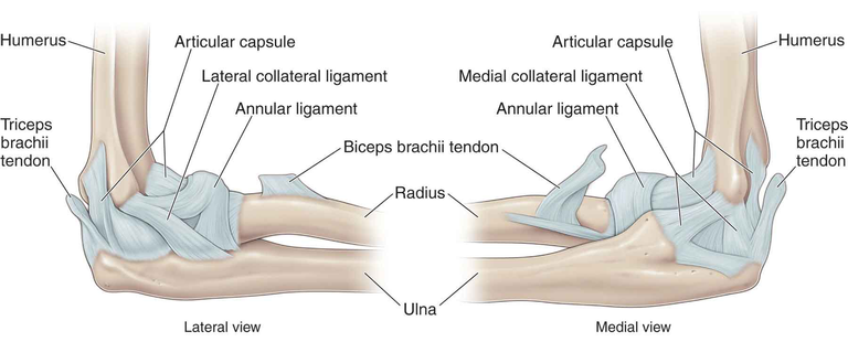
The proximal radioulnar joint is stabilized by several crucial ligaments that help maintain the integrity of the joint and support its range of motion. The key ligaments involved in the proximal radioulnar joint include the annular ligament, the quadrate ligament, and the accessory ligaments.
Annular Ligament:
- The annular ligament encircles the head of the radius, holding it in place within the radial notch of the ulna.
- This ligament allows for smooth rotation of the forearm during pronation and supination while preventing the radius from dislocating from the joint.
- It is a strong, fibrous band that plays a central role in stabilising the proximal radioulnar joint and facilitating its pivotal movements.
Quadrate Ligament:
- The quadrate ligament attaches the neck of the radius to the ulna, providing additional stability to the proximal radioulnar joint.
- This ligament complements the function of the annular ligament in maintaining the alignment of the radius and ulna bones during forearm rotation.
- It helps prevent excessive movement and ensures that the joint can withstand the forces applied during everyday activities.
Accessory Ligaments:
- In addition to the annular and quadrate ligaments, the proximal radioulnar joint is supported by accessory ligaments that contribute to its stability and function.
- These ligaments vary in size and location but work together to reinforce the joint capsule and prevent excessive movement that could lead to injury.
- While not as prominent as the annular and quadrate ligaments, these accessory ligaments play a significant role in ensuring the overall strength and resilience of the proximal radioulnar joint.
The coordination of these ligaments is important for maintaining joint health and functionality, reinforcing the importance of proper care and maintenance of this anatomical structure.
Movements
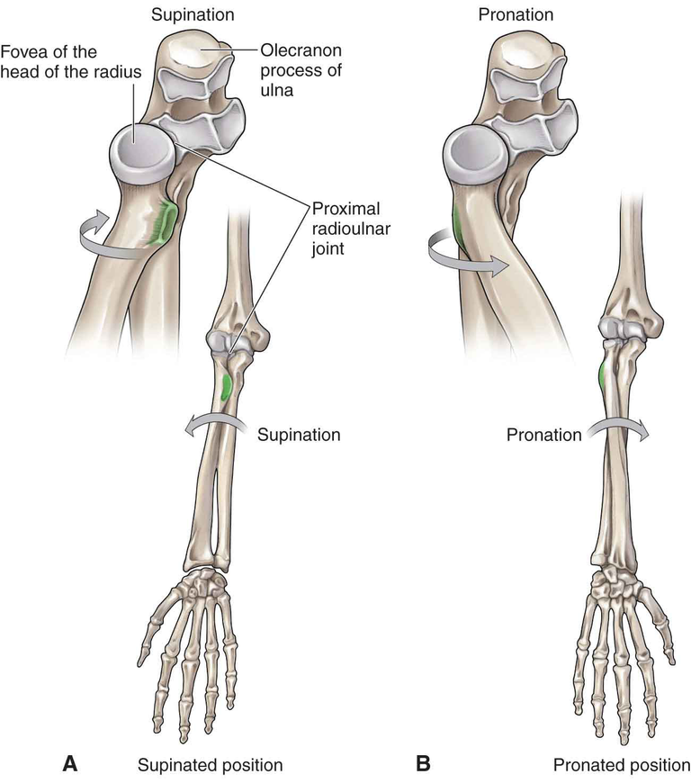
The proximal radioulnar joint is a pivotal joint in the forearm that allows for essential movements like pronation and supination. These movements are facilitated by the articulation between the head of the radius and the radial notch of the ulna, along with the supporting ligaments and muscles surrounding the joint.
Pronation:
- Pronation is the motion that rotates the palm of the hand to face downward or backward.
- During pronation, the radius rotates over the ulna, moving from a position where the palm faces up (supine) to a position where the palm faces down (pronated).
- The annular ligament plays a crucial role in stabilizing the proximal radioulnar joint during pronation, allowing for smooth and controlled movement of the forearm.
Supination:
- Supination is the opposite motion of pronation, rotating the palm of the hand to face upward or forward.
- In supination, the radius returns to its neutral position alongside the ulna, with the palm facing up.
- The quadrate ligament, along with other supporting ligaments and muscles, aids in stabilizing the joint during supination and ensuring proper alignment of the radius and ulna.
Coupled Motion:
- Pronation and supination occur as a coupled motion, meaning that when one motion is activated, the other is passively engaged.
- The annular ligament allows the radius to glide smoothly over the ulna during both pronation and supination, maintaining joint stability and facilitating coordinated movement of the forearm.
Range of Motion:
- The proximal radioulnar joint has a significant range of motion, allowing for approximately 180 degrees of pronation and supination.
- This wide range of motion is crucial for everyday activities such as turning a doorknob, using utensils, or typing on a keyboard, highlighting the importance of a healthy and functional proximal radioulnar joint.
Muscular Involvement
Muscles surrounding the proximal radioulnar joint play a vital role in initiating and controlling pronation and supination movements.
Muscles such as the pronator teres and pronator quadratus are responsible for pronation, while muscles like the biceps brachii and supinator facilitate supination.
The coordinated action of these muscles, along with the ligaments and joint structures, ensures smooth and efficient movement of the forearm during pronation and supination.
Overall, the movement of the proximal radioulnar joint does a nice job when it comes to maintaining functional mobility in the forearm and upper extremity. If we can understand the anatomy and mechanics of this joint, we'll be provided with the insight into the structures and muscles that enable us to perform a wide range of tasks in our daily lives.
Blood Supply and Innervation
The blood supply and innervation of the proximal radioulnar joint play key roles in ensuring its function and providing sensation to the surrounding structures.
Blood Supply
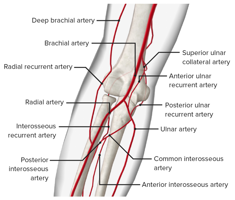
The blood supply to the proximal radioulnar joint primarily comes from branches of the radial and ulnar arteries.The radial recurrent artery, a branch of the radial artery, provides blood to the lateral aspect of the joint, while the ulnar recurrent artery, a branch of the ulnar artery, supplies the medial aspect.
These arteries branch into smaller vessels that form a rich network of blood supply around the joint, ensuring adequate oxygen and nutrients reach the joint structures and surrounding tissues.
Proper blood supply is essential for maintaining the health and function of the joint, as well as supporting the repair and regeneration of tissues in case of injury or damage.
Innervation
The proximal radioulnar joint receives innervation from branches of the radial nerve and the anterior interosseous nerve.
The radial nerve gives rise to branches that provide sensory innervation to the lateral aspect of the joint, while the anterior interosseous nerve innervates the anterior aspect.
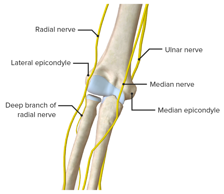
Sensory nerves in the joint play a critical role in relaying information about position, movement, and pain to the central nervous system, allowing for proprioception and protective responses.
In addition to sensory innervation, motor branches of these nerves also supply muscles that act on the proximal radioulnar joint, contributing to its stability and movement control.
Clinical Implications:
- Damage or injury to the blood supply or nerves innervating the proximal radioulnar joint can result in pain, altered sensation, and impaired function.
- Vascular injuries may compromise the blood flow to the joint, leading to impaired healing, increased risk of infection, and reduced overall joint health.
- Nerve injuries can result in sensory deficits, muscle weakness, and coordination problems affecting the movement and stability of the joint.
- Proper assessment and management of vascular and nerve issues are essential in addressing potential complications and ensuring optimal recovery and function of the proximal radioulnar joint.
Note, refer to your Atlas textbooks or online pictures while reading this article. This will help you to catch up and understand fully what the article is all about
Finally, this is the end of the article. I hope you enjoyed the descriptions and as well acknowledge why you are able to move your foreman in those complicated ways!!!
See you soon
References
(1) Proximal radioulnar joint: Anatomy, movements | Kenhub. https://www.kenhub.com/en/library/anatomy/proximal-radioulnar-joint.
(2) The Radioulnar Joints - TeachMeAnatomy. https://teachmeanatomy.info/upper-limb/joints/radioulnar-joints/.
(3) Proximal radioulnar articulation - Wikipedia. https://en.wikipedia.org/wiki/Proximal_radioulnar_articulation.
(4) en.wikipedia.org. https://en.wikipedia.org/wiki/Proximal_radioulnar_articulation.


Thanks for your contribution to the STEMsocial community. Feel free to join us on discord to get to know the rest of us!
Please consider delegating to the @stemsocial account (85% of the curation rewards are returned).
You may also include @stemsocial as a beneficiary of the rewards of this post to get a stronger support.