Simplifying the Anatomy of the upper limb: Muscles of the Shoulder Region
Introduction
Hey friends, last week, we discussed the arterial blood supply of the upper limb and today, we'll be looking at a different thing although related to the upper limb.
Yes, you can't discuss a particular region of the body without mentioning the muscles that enable it to function properly.
The shoulder region is important for helping us move our arms and stay stable. Knowing about the muscles in this area can give us insight into how our arms work. In this article, we're going to take a closer look at the main muscles in the shoulder region, where they come from, where they attach, and how they get their blood and nerve supply.
So what are we waiting for? Let's dive into the anatomy and see how these muscles team up to support our arm movements!
Deltoid Muscle
The deltoid muscle is a powerhouse in the shoulder region, allowing us to perform a wide range of arm movements with strength and precision. Its structure and function help us in maintaining shoulder stability and enabling us to engage in various everyday and athletic activities.
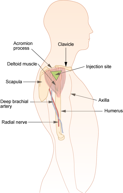
The deltoid muscle is known for its distinctive triangular shape that covers the shoulder joint.
Origin: The deltoid muscle originates from three main points - the clavicle, the acromion process of the scapula, and the spine of the scapula. These origins provide a broad base for the muscle to arise from.
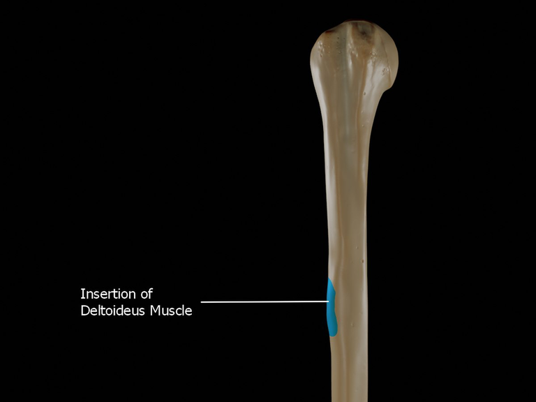
Insertion: The deltoid muscle inserts at the deltoid tuberosity of the humerus, the bony prominence on the lateral side of the upper arm bone. This insertion point allows the deltoid muscle to exert its pulling or lifting action on the arm.
Function: The deltoid muscle is responsible for several crucial arm movements, including abduction (raising the arm away from the body), flexion (bringing the arm forward), and extension (moving the arm backward). It helps in various daily activities like reaching overhead or throwing a ball.
Blood Supply: The blood supply to the deltoid muscle comes from the posterior circumflex humeral artery, ensuring that the muscle receives oxygen and nutrients for its functions.
Nerve Supply: The deltoid muscle is innervated by the axillary nerve, which originates from the brachial plexus (specifically from the C5 and C6 nerve roots). This nerve provides the necessary signals for the muscle to contract and move the arm effectively.
Do you wish to improve your deltoid muscle, then hit the gym!!
The Pectoral Muscles
The pectoral muscles, which are key muscles of the chest region have a significant role in various arm movements and shoulder stability.
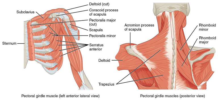
Pectoralis Major Muscle:
- Origin: The Pectoralis major muscle has two heads - a clavicular head that originates from the medial half of the clavicle, and a sternocostal head that originates from the sternum, upper six costal cartilages, and the aponeurosis of the external oblique muscle.
- Insertion: It inserts onto the lateral lip of the intertubercular groove of the humerus, providing strength for arm movements.
- Function: The Pectoralis major muscle is responsible for medially rotating, flexing, and adducting the arm. It also plays a role in stabilizing the shoulder joint during various movements.
- Blood Supply: The muscle receives its blood supply from branches of the thoracoacromial artery, ensuring good vascularization.
- Nerve Supply: It is innervated by the medial and lateral pectoral nerves, which are branches of the brachial plexus coming from the C5 to T1 nerve roots.
Pectoralis Minor Muscle:
- Origin: The Pectoralis minor muscle originates from the third, fourth, and fifth ribs near their cartilages.
- Insertion: It inserts onto the coracoid process of the scapula, aiding in the stabilization and movement of the scapula.
- Function: The Pectoralis minor muscle plays a role in protracting and depressing the scapula, assisting in movements involving the shoulder girdle.
- Blood Supply: Blood supply to the muscle is provided by branches of the thoracoacromial artery, ensuring proper circulation.
- Nerve Supply: It is innervated by the medial pectoral nerve, a branch of the brachial plexus originating from the C8 and T1 nerve roots.
Subscapularis Muscle
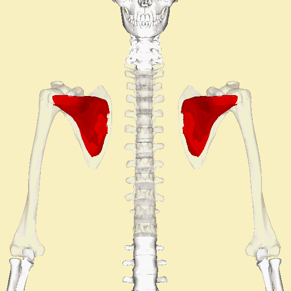
The subscapularis muscle is another great muscle located on the anterior (front) side of the shoulder joint. This muscle has important features which we will be looking at in a moment. Let's begin with its origin.
Origin: The subscapularis muscle originates from the subscapular fossa, a concave depression on the anterior aspect of the scapula (shoulder blade). It has a broad attachment area that provides a strong foundation for its functions.
Insertion: The muscle inserts onto the lesser tubercle of the humerus, a bony prominence on the front of the humerus bone. This insertion point allows the subscapularis muscle to exert its actions on the shoulder joint.
Function: The subscapularis muscle exhibits its action in medially rotating the arm (turning the arm inward) and stabilising the shoulder joint. It also helps in preventing dislocation of the humeral head during arm movements.
Blood Supply: The blood supply to the subscapularis muscle is mainly provided by branches of the subscapular artery, ensuring that the muscle receives the necessary nutrients and oxygen for its function.
Nerve Supply: The subscapularis muscle is innervated by the upper and lower subscapular nerves, which are branches of the brachial plexus arising from the C5 to C7 nerve roots. These nerves provide the muscle with the necessary signals to contract and perform its actions.
Again, I'll like you to note that the subscapularis muscle plays a significant role in the stability and movement of the shoulder joint, particularly in medially rotating the arm and preventing dislocations.
The Teres Muscles
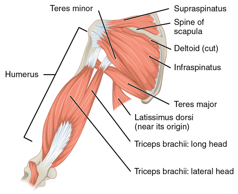
These are two significant muscles located at the back of the shoulder region. Their functions and importance can never be overlooked especially when it comes to how we move our shoulder joint.
Teres Major Muscle:
Origin: The Teres major muscle originates from the inferior angle and lower part of the lateral border of the scapula, providing a sturdy base for its actions.
Insertion: It inserts onto the medial lip of the intertubercular groove of the humerus, near the insertion point of the Latissimus dorsi muscle.
Function: The Teres major muscle assists in extending, medially rotating, and adducting the arm. It works in coordination with other shoulder muscles to facilitate various arm movements.
Blood Supply: The Teres major muscle receives its blood supply from the subscapular artery, ensuring proper nourishment for optimal function.
Nerve Supply: It is innervated by the lower subscapular nerve, a branch of the brachial plexus originating from the C5 and C6 nerve roots.
Teres Minor Muscle:
Origin: The Teres minor muscle originates from the lateral border of the scapula, specifically from the upper two-thirds of this border.
-Insertion: It inserts onto the greater tubercle of the humerus, adjacent to the insertion points of the Infraspinatus muscle.
Function: The Teres minor muscle primarily assists in the external rotation of the arm and helps stabilize the shoulder joint during arm movements.
Blood Supply: Blood supply to the Teres minor muscle is provided by the posterior circumflex humeral artery, ensuring proper circulation for its functions.
Nerve Supply: The Teres minor muscle is innervated by the axillary nerve, a branch of the brachial plexus arising from the C5 and C6 nerve roots.
Recognise that while the Teres major contributes to arm adduction, extension, and medial rotation, the Teres minor focuses on arm external rotation and shoulder joint stability. Together with other shoulder muscles, they enable various arm movements and support the overall function of the shoulder complex.
Subclavius Muscle
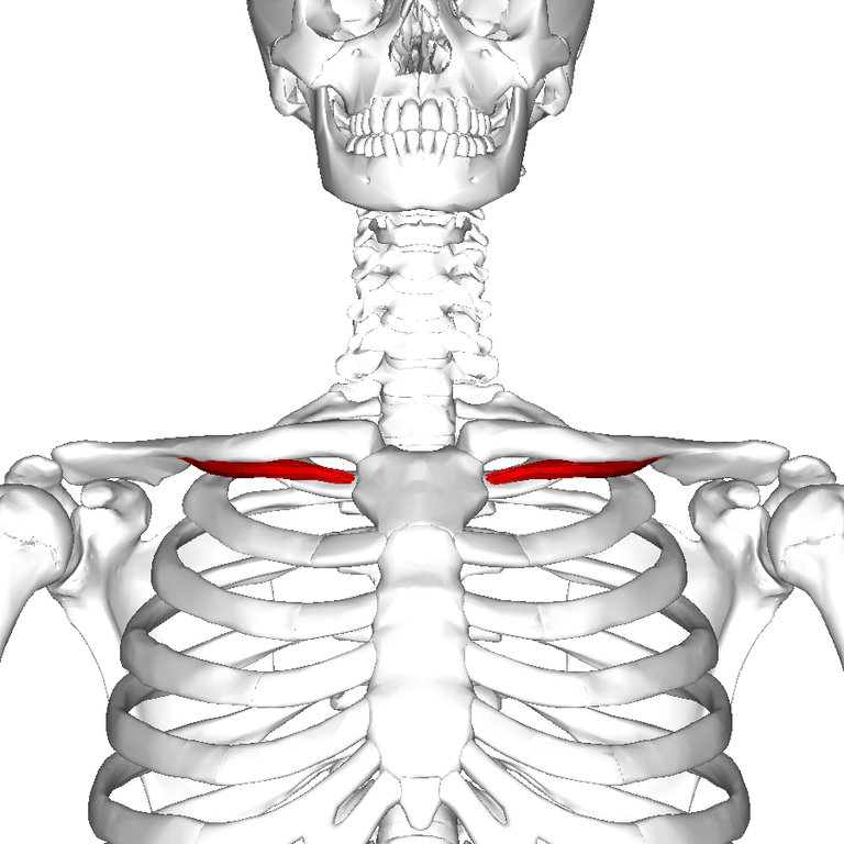
The subclavius muscle is a small yet significant muscle located beneath the clavicle (collarbone) that plays a role in stabilizing the shoulder joint and assisting with certain arm movements. Here's an overview of the subclavius muscle:
Location: The subclavius muscle is a thin, triangular muscle situated beneath the clavicle, running from the first rib to the undersurface of the middle portion of the clavicle. It lies deep to the clavicular head of the pectoralis major muscle.
Function:
Stabilization: The subclavius muscle helps stabilize the clavicle by preventing excessive movement or elevation of the bone during arm movements, especially overhead motions.
Protection: It provides protection to the underlying neurovascular structures (brachial plexus and subclavian vessels) in the region.
Supportive Role: While the subclavius muscle itself may not be directly involved in producing significant movements, its stabilizing function is crucial for maintaining proper shoulder mechanics and preventing injuries.
Nerve Supply: The subclavius muscle receives its nerve supply from the subclavius nerve, a branch of the superior trunk of the brachial plexus (C5-C6 spinal nerve roots). This innervation allows for controlled activation of the muscle.
Clinical Significance: Injuries to the subclavius muscle are rare but can occur in cases of direct trauma to the area or repetitive stress. Such injuries may result in pain, weakness, or altered shoulder mechanics, necessitating appropriate evaluation and management.
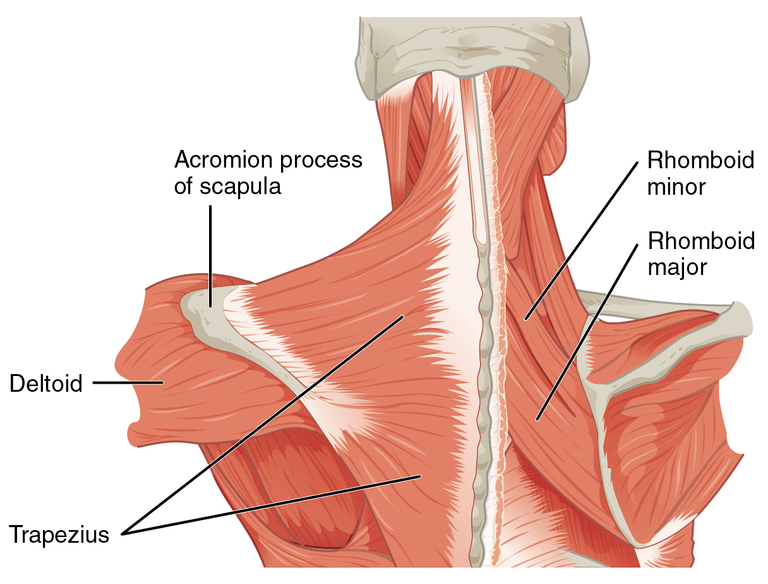
Trapezius

The trapezius muscle, commonly referred to as the "traps," is a large, powerful muscle in the upper back and neck region. Here's an overview of the trapezius muscle:
Location: The trapezius muscle is a flat, triangular muscle that extends from the base of the skull down to the middle of the back and laterally to the shoulders. It covers a significant portion of the upper back and neck area.
Structure: The trapezius muscle is divided into three main parts: the upper fibers (descending from the occipital bone), the middle fibers (running horizontally from the spine to the scapula), and the lower fibers (ascending from the spine to the scapula).
Functions
Upper fibers: Elevate the shoulders (e.g., shrugging motions) and support movements like raising the arms overhead.
- Middle fibers: Retract the scapulae (pull the shoulder blades toward the spine) during movements like rowing or pulling motions.
Lower fibers: Depress the scapulae (lower the shoulder blades) and assist in maintaining proper posture.
Activities: The trapezius muscle is heavily involved in various daily activities and exercises, including shoulder shrugs, upright rows, pulling movements (rows, pull-ups), and activities requiring scapular stabilization.
Nerve Supply: The trapezius muscle is innervated by the spinal accessory nerve (cranial nerve XI) and branches of the cervical spinal nerves (C3-C4). This innervation allows for voluntary control over the muscle's movements.
References
(1) Muscles of the Shoulder Region - TeachMeAnatomy. https://teachmeanatomy.info/upper-limb/muscles/shoulder/.
(2) Shoulder muscles : Anatomy and functions | Kenhub. https://www.kenhub.com/en/library/anatomy/shoulder-muscles.
(3) Shoulder Anatomy, Area & Diagram | Body Maps - Healthline. https://www.healthline.com/human-body-maps/shoulder.
(4) The Extrinsic Muscles of the Shoulder - TeachMeAnatomy. https://teachmeanatomy.info/upper-limb/muscles/shoulder/extrinsic/.
(5) Shoulder Muscles: Anatomy, Function & Common Conditions - Cleveland Clinic. https://my.clevelandclinic.org/health/body/21798-shoulder-muscles.
(6) Getty Images. https://www.gettyimages.com/detail/photo/young-woman-pain-left-shoulder-ache-in-human-body-royalty-free-image/1159624713.


I am a complete beginner who resides in Africa's Western Hemisphere. My name is James, but you may reach out to me through the Facebook page [James Kossy] (https://www.facebook.com/christ.messenger.904) Physics, chemistry, and biology are the three topics that I find most enjoyable. My current studies are taking place at the university level, with the intention of becoming a recognized professional in physiotherapy. I am fascinated by all things technological, and I take pleasure in contributing to the fascinating technological advancements that are taking place throughout the world today. In my spare time, I'd like to learn more about programming and help others with any technical problems they may be having. 💞 ***🌹❤️ Thank you so much to everyone who has supported me thus far. ****💞 At the moment, I don't have the right words to say how much I appreciate all of your help. You never cease to astonish me with your generosity. For me, this has turned into a haven of enjoyment. Thanks to colleagues like you, this has all been possible. You've been a great support for me. Everything you have done for me and my family has been greatly appreciated, and I will always be grateful to you. 💕.
Posted Using InLeo Alpha
!LOLZ
lolztoken.com
He tells her about his job.
Credit: marshmellowman
@jsalvage, I sent you an $LOLZ on behalf of benefice-net
(7/10)
Delegate Hive Tokens to Farm $LOLZ and earn 110% Rewards. Learn more.
Thank so much fren
Hello.
There is reasonable evidence that this article is machine-generated.
We would appreciate it if you could avoid publishing AI-generated content (full or partial texts, art, etc.).
Thank you.
Guide: AI-Generated Content = Not Original Content
If you believe this comment is in error, please contact us in #appeals in Discord.
Thanks for your contribution to the STEMsocial community. Feel free to join us on discord to get to know the rest of us!
Please consider delegating to the @stemsocial account (85% of the curation rewards are returned).
You may also include @stemsocial as a beneficiary of the rewards of this post to get a stronger support.
what if all ligaments in the shoulder are ripped?
and the collarbone stands on the height of the ear?
Acromioclavicular joint fusion on top of that
and now some artificial carbon band, after years of chronification, but at least collarbone not on ear height anymore and not a complete cripple anymore
but permanent pain and hard cramps
but probably "normal" ?
especially with such bad medicine (didnt even have post-treatment, whole shoulder was frozen then..)
Wow a very well detailed explanation. You really invested quite a lot of effort in making this out and it actually comes out so nice
Thank you so much for your time, it is greatly appreciated
Dear @jsalvage, great health article as always. It's great to have you on Hive and see your journey here. Have a great day, zuerich
Thank you so much for stopping by. And indeed, I had a wonderful day today although. Many things to write home about !