Cranial Meninges - Neuroanatomy
It's time to delve into the realm of brain anatomy! Today, we'll examine the meninges, a vital defense system that encircles the brain. Even though it sounds like a technical term, knowing about the meninges will help us to understand the brain's complex functions and preserving its health.
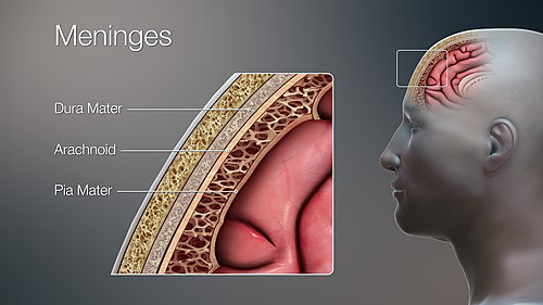
Consider the meninges as the brain's own three-layered bodyguard, protecting the delicate and priceless organ known as our brain. These layers—also referred to as the dura mater, arachnoid mater, and pia mater—are what maintain the security and ideal operation of the brain.
Why is it necessary to understand meninges? Now picture a sturdy stronghold encircling a priceless loot. Similar insulation against possible damage is provided to the brain by the meninges. Understanding the meninges' anatomy helps us recognize problems that may compromise the health of the brain and provides insight into its weaknesses.
By the time you finish reading this essay, you will have a better understanding of these amazing layers as well as how they advance our knowledge of brain health and related disorders.
The Dura Mater
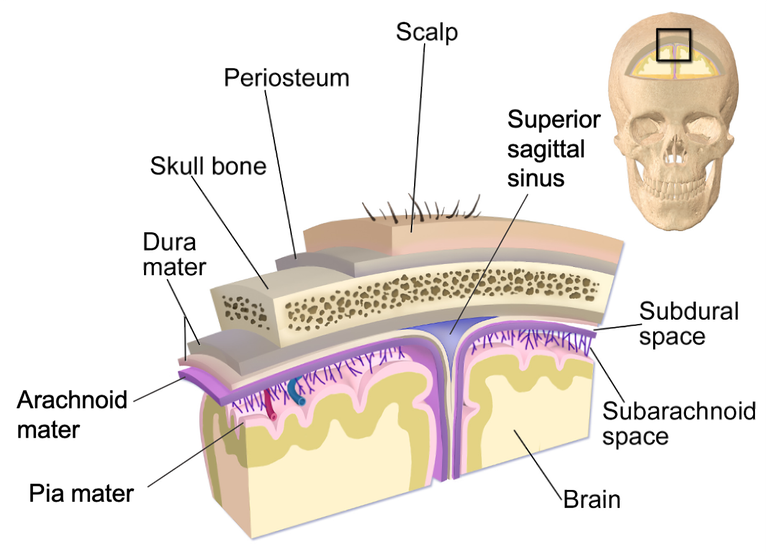
The brain and spinal cord are encased in a strong, fibrous membrane called the dura mater, which is the outermost and hardest layer of the meninges. The Latin term "dura mater," which means "tough mother" or "hard mother," accurately describes its strong and protective character.
Organization
The periosteal layer and the meningeal layer are the two separate layers that make up the dura mater. The dura mater and the bone are strongly demarcated by the periosteal layer, which firmly adheres to the inside surface of the skull. The arachnoid mater, the layer underlying the periosteal layer, is firmly adhered to by the meningeal layer, which is situated deep within it.
Divisions
There are two primary divisions of the dura mater within the cranium, or skull:
Endosteal Dura: The dura mater's outermost division is known as the endosteal dura. It forms a barrier of defense between the brain and the bone along the inside surface of the skull. In comparison to the inner meningeal layer, this layer is comparatively thicker and less vascular.
Meningeal Dura: Adhering tightly to the arachnoid mater, the meningeal dura is the innermost division located underneath the endosteal dura. Compared to the endosteal dura, it is more sensitive and thinner. The meningeal dura forms different structures inside the skull as it spreads into the cranial cavity. It creates the falx cerebri, a fold with a sickle shape that extends into the longitudinal fissure that divides the two hemispheres of the brain. The meningeal dura also forms the diaphragma sellae, a sheet covering the pituitary gland inside the sella turcica of the sphenoid bone, and the tentorium cerebelli, a fold dividing the cerebellum from the cerebrum.
Functions
The brain and spinal cord are vitally protected by the dura mater. Its robust and fibrous structure serves as a barrier, preventing shock and mechanical harm to these fragile components. In addition to providing protection, the dura mater aids in maintaining the brain's correct location within the skull by stabilizing and supporting it.
Arachnoid
The middle layer of the meninges, the arachnoid mater, is situated between the dura and pia maters. Its name comes from the way it looks—like the web of a spider. Let's take a closer look at its composition and purpose.
Structure
Made of fibrous connective tissue, the arachnoid mater is a thin, delicate membrane. It is comparatively avascular since it is devoid of blood vessels. The subdural space is formed by the contact between the inner surface of the dura mater and the outer surface of the arachnoid layer. The subarachnoid space is formed by the tight application of the arachnoid layer's inner surface to the pia mater. Cerebrospinal fluid (CSF) is found in the subdural and subarachnoid regions.
Protecting the brain and spinal cord underneath is the primary purpose of the arachnoid mater. It physically divides the fragile brain tissue from the dura mater above and the pia mater below, serving as a barrier. This division lessens the possibility of harm or injury by preventing direct contact between the fragile neural tissue and the hard bone.
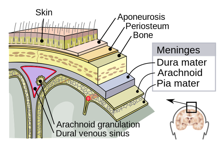
Cerebrospinal fluid (CSF) circulation and absorption is another important function of the arachnoid mater. Arachnoid granulations, also known as villi, are structures found in the arachnoid layer that are tiny protrusions into the superior sagittal venous sinus, a major vein found in the dura mater. These granulations enable the central nervous system's fluid balance to be continuously maintained by allowing the CSF to be absorbed into the venous system.
The pia mater
The deepest layer of the meninges, the pia mater, is situated in closest proximity to the brain and spinal cord. It is a thin, highly vascularized membrane that is essential for maintaining and nourishing the brain tissue underneath. Let's take a closer look at its composition and purpose.
Structure
Adhering tightly to the surface of the brain and spinal cord is the thin, translucent pia mater membrane, which is made of delicate connective tissue. It provides total coverage by following the morphology of the neural tissue, covering the gyri (ridges) and sinking into the sulci (grooves) of the brain. Due to its high vascularization, the pia mater has an extensive blood artery network that supplies the brain tissue with nutrients and oxygen.
Protecting and supporting the sensitive neuronal tissue of the brain and spinal cord is the pia mater's main purpose. It protects the brain tissue from mechanical damage by functioning as a barrier between it and the surrounding structures.
Furthermore, the pia mater is essential for the central nervous system's nutrition. The underlying neurons and glial cells receive oxygen, glucose, and other vital nutrients from the blood arteries within the pia mater. The synthesis of energy and the general health of the neural tissue depend on these nutrients.
Moreover, the pia mater plays a role in the synthesis and movement of cerebrospinal fluid (CSF), which supports and shields the brain and spinal cord. The pia mater sends tiny projections into the subarachnoid region that resemble fingers and are known as pial villi. In order to maintain CSF volume and pressure, these villi permit CSF to move between the blood arteries and the subarachnoid space.
Common meninges-related illnesses or ailments
Meningitis
Meningitis is an inflammation of the meninges that can be brought on by infections with bacteria, viruses, or fungi. There are several ways that the infection might enter the meninges, including the circulation or respiratory droplets.
Intracranial hemorrhage
Bleeding that takes place in the space between the meninges' arachnoid and pia mater layers is referred to as a subarachnoid hemorrhage. This kind of bleeding frequently happens as a result of a head injury or a ruptured cerebral aneurysm.
Symptoms and Available Therapies
First, meningitis
Symptoms: Meningitis symptoms include intense headaches, stiff necks, elevated fever, photosensitivity, nausea, vomiting, and altered mental status. In addition, irritability, inadequate eating, and a protruding fontanelle may be seen in babies.
Diagnosis: A lumbar puncture (spinal tap), which includes examining the cerebrospinal fluid for evidence of infection, is one of several diagnostic tests used in conjunction with a comprehensive physical examination, symptom analysis, and other diagnostic procedures.
Treatment: Meningitis needs to be seen by a doctor right away. In order to cure the underlying illness, treatment frequently entails hospitalization for intravenous antibiotics or antiviral drugs. It's also critical to provide supportive care, which includes pain relief and hydration balance.
Hemorrhage Subarachnoid
Symptoms: A subarachnoid hemorrhage can cause a variety of symptoms, such as a sudden, intense headache that is frequently referred to as the worst headache of one's life, stiff neck, nausea, vomiting, light sensitivity, seizures, and loss of consciousness.
Diagnosis: Making a diagnosis entails a thorough assessment that includes a physical examination, a study of the patient's medical history, and imaging tests such an MRI or CT scan.
Treatment: The course of action is determined by the extent and underlying reason of the hemorrhage. In order to stabilize the patient's condition, it may involve supportive care, surgical procedures such aneurysm coiling or clipping, and drugs to control symptoms and avoid complications.
Subarachnoid hemorrhages and meningitis are both dangerous illnesses that need to be treated right away. Timely identification and treatment are required in order to reduce long-term damage and avoid problems.
Apart from the therapies already discussed, symptom management and supportive care are crucial elements of the comprehensive treatment strategy. This could entail managing pain, taking anti-inflammatory drugs, keeping an eye on vital signs, and assessing neurological state.
Another important factor is prevention, especially in cases of bacterial meningitis. There are vaccinations available against a number of bacterial types that might result in meningitis, including Streptococcus pneumoniae, Neisseria meningitidis, and Haemophilus influenzae type b (Hib). Meningitis can be prevented by maintaining current immunization records, washing your hands frequently, and avoiding close contact with those who are sick.
References
- https://www.ncbi.nlm.nih.gov/books/NBK539882/
- https://www.kenhub.com/en/library/anatomy/meninges-of-the-brain-and-spinal-cord
- https://teachmeanatomy.info/neuroanatomy/structures/meninges/
- https://my.clevelandclinic.org/health/articles/22266-meninges#:~:text=Your%20brain%20and%20spinal%20cord%20are%20protected%20and%20supported%20by,as%20well%20as%20other%20functions.
- https://www.webmd.com/brain/meninges-what-to-know
- https://www.mayoclinic.org/diseases-conditions/meningioma/multimedia/meninges/img-20008665
- https://study.com/learn/lesson/meninges-layers-function-anatomy.html


Thanks for your contribution to the STEMsocial community. Feel free to join us on discord to get to know the rest of us!
Please consider delegating to the @stemsocial account (85% of the curation rewards are returned).
You may also include @stemsocial as a beneficiary of the rewards of this post to get a stronger support.