The Anatomy, and Physiology of the Ear
It is no doubt that all of our sense organs are important to us for certain special abilities, such as sight (eyes), ears for sound, nose for smell, tongue for taste, and skin for touch, but I was having a conversation with my friend on Sunday, when we went for lunch, and in the middle of our conversation, she said that her ears are one of the sense organs that she really do not know existed unless she wants to clean wax from it. She went further to say she really does not pay attention to her ears, it just functions, unlike the eyes where she always wants to protect, or her tongue that she cares for regularly. I told her in simple words, "All senses are important, you should treat them with utmost respect".
I will be doing a series on the Sense Organs in the body, and today, I will be starting with the ear. Furthermore, I will be explaining the anatomy, and physiology of the ear in this post. So let's begin the hearing process
Did you say hearing process? Okay! I can hear you, and I will be hearing you/listening to you.
The Anatomy of the Outer Ears
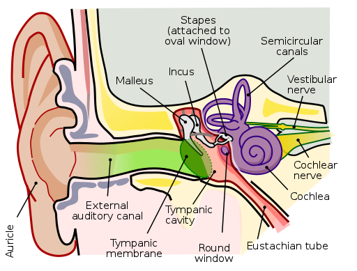
The ear is divided into three part, the external, the middle, and the inner part of the ear. The external part of the ear, starts with the Pinna/Auricle, which is held by the External Acoustic Meatus that divides the external ear from the middle ear. The Pinna is the outermost part of the ear, which is covered with squamous epithelial tissue. The outermost of the skin is made up of squamous epithelial tissue, which prevents the skin from bacteria and friction. Just inside the Pinna, is the elastic cartilage. The elastic cartilage can be found in a few parts of the body including the ear, the nose, and the epiglottis, and the purpose is to help the part of the body it is in to be able to stretch and retract back to its position. In the Pinna is the auriculotemporal branch, which provides sensory innervation to the External Acoustic Meatus. Immediately after the Pinna is a hole that leads to the middle and inner ear. You should see this hole in your ear, that is the External Acoustic Meatus. In the External Acoustic Meatus is the Ceruminous glands which produces the ear wax (cerumen).
I am sure you must have cleaned those ears wax in your ear, just like my friend does, and almost everyone does. The ear wax is produced by the Ceruminous glands
The Cerumen is important for protecting the inner part of the ear. It protects the inner ear from both physical invasions (insects), and microbial invasion. Immediately after the External Acoustic Meatus is the Tympanic Membrane, literally known as the eardrum, made up of the Pars Tensa, and the Pars Flaccida. It separates the outer ear from the middle ear, and vibrates when sound waves reaches it.
In the process of cleaning earwax in the ear, inserting objects too deep into the ear could puncture the Pars flaccida. You know that scenario when you hear of a person having a burst eardrum? That's the case.
The Anatomy of the Middle Ear
Just Behind the Tympanic Membrane is the middle ear, where tiny bones called the ear ossicles are located. The bones are the Malleus, Incus, and Stapes.
The Tensor Tympani is a muscle connected to the Malleus, helping to reduce/dampen the amplitude of sound vibration entering into the inner part of the ear. The Malleus is connected to the Incus, and the Incus is connected to the Stapes. Sound waves from the outer ear gets to the tympanic membrane, and vibrates the tiny bones in the middle ear called the ossicles. The malleus will be vibrated, which will then vibrate the incus then the stapes, which then sends the vibration to the oval window.. The Oval Window gets the vibration and turns into a fluid filled vibration in the Cochlea..
Still in the middle ear, is the Stapedius Muscle, which is the smallest skeletal muscle in the body. It dampens the effect of the Stapes to the oval window. The Stapedius Muscle is controlled by the parasympathetic fibers, and the facial nerve also known as the Seventh Cranial nerve.. Branching from the facial nerve that connects to the stapedius muscle is the Chorda Tympani. The Chorda Tympani runs from the lingual nerve in the taste bud, and run through the middle ear. The Chorda tympani relays sensations of taste to the brain.
In the Medial wall of the meddle ear, the Cochlea presses in from the internal wall, creating the Cochlea promontory. On the Tympanic promontory, the tympanic plexus is formed from branches of Jacobson's nerve (glossopharyngeal nerve). Still in the middle ear, the faringial tympanic tube (Eustachian tube), is a tube that connects the middle ear to the lateral wall of the nasopharynx. It equalizes the pressure of air within the ear and the atmospheric pressure. At the posterior part of the middle ear is the Mastoid which also does the process of air maintaning..
The Anatomy of the Inner Ear
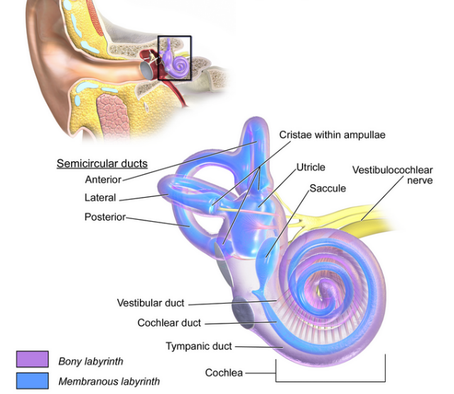
The inner ear isn't started without mentioning the cochlea. The Cochlea is divided into three chambers which are the Scala vestibuli, Scala Media/cochlea duct, and the Scala Tympani. The Scala Vestibuli is the upper chamber of the Cochlea, which comes immediately after the oval window. The Scala Vestibuli is made up of fluid called the perilymph. The Scala Tympani is also made up of the perilymph, and it is located close to the Round window membrane, which is inferior to the oval window. The apex point in the shell-like structure of the inner ear, where the Scala Vestibuli, and the Scala Tympani meets is known as the Helicotremo. The Scala media is made up of a fluid called the endolymph (inner membranous labyrinth).
In the inner ear, there is a branch from the clochlea, known as the Vestibulocochlea, which consists of the vestibular and cochlear nerves. Connecting the Cochlea and the semicircular canal of the ear to help maintain static equilibrium of sound while moving against the sound vibration, is the Vestibule, which is made up of two membranous labyrinths called the Utricle and the Saccule.
The semicircular canal is made up of the anterior, posterior, and the lateral canal. It is the outer bony labyrinth made up of perilymph. The semicircular canals are dilated at the Ampulla. The Ampulla is contains of sensory epithelium which is made up of hair cells, responsible for dynamic equilibrium (such as head rotation).
Conclusion
The ear is made up of three parts. The external ear, which is the Pinna/auricle made up of elastic catillages. This is the part you can touch and move with your hands. The external ear is connected to the middle ear through the External Accoustic Meatus. The middle ear and external is divided by the Tympanic Membrane. The Tympanic Membrane is the eardrum. and it seperats the middle ear bones (ossicles) from the external ear. the middle ear is seperated from the inner ear via he Oval Window. The Oval window seperates the Cochlea which is the major part of the inner ear from the middle ear. The ear is responsible to converting and sending sound waves to the brain to be interpreted, they help to maintain air pressure which enters the ear, and maintain equilibrum.
Image Reference
Image 1 || Wikimedia Commons
Thanks for this very clear description, and all the references for the technical words. This time, I have no specific comment or question (everything was clear), and only wanted to show that I have read this post (that has now a first comment ;) ).
Thanks a lot @lemouth for the comment.. This means so much to me. I must confess that I feel elated by your comments. Thanks a lot for always stopping by.
You are very welcome!
Thanks for your contribution to the STEMsocial community. Feel free to join us on discord to get to know the rest of us!
Please consider delegating to the @stemsocial account (85% of the curation rewards are returned).
Thanks for including @stemsocial as a beneficiary, which gives you stronger support.