Normal Thyroid vs Papillary Thyroid Carcinoma
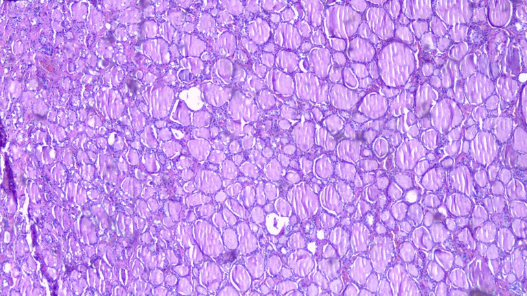
The following three images are the normal architecture of the thyroid gland and then followed up by a case of papillary thyroid carcinoma. Thyroid tissue is made up follicles containing colloid and lined by simple cuboidal epithelium. If you recall your biology class, cuboidal cells are cuboids in shape and the form associated with glands. The cells have round, normochromatic nuclei with inconspicuous nucleoli and moderate cytoplasm.
Taken at Scanner View (40x)
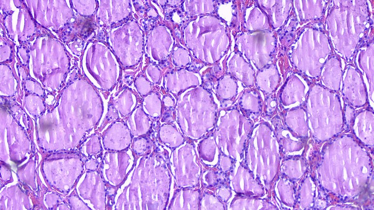
Taken at Low Power View (100x)
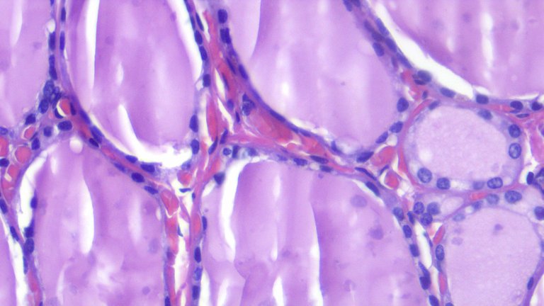
Taken at High Power View (400x)
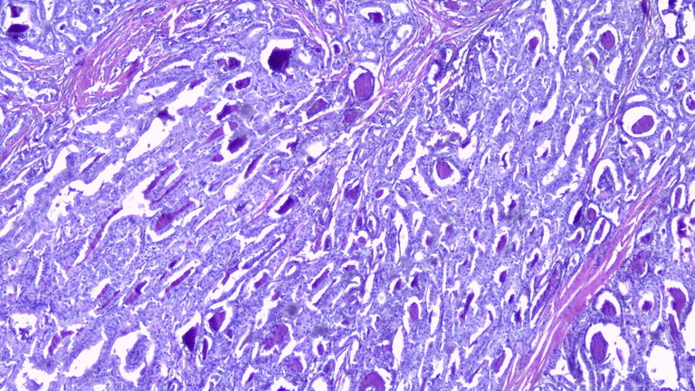
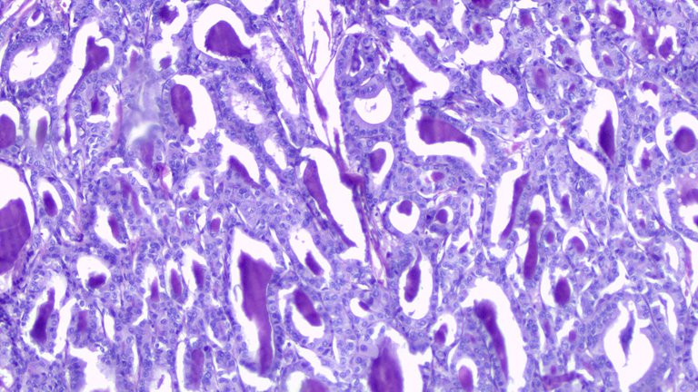
Diagnosis of Papillary Thyroid Carcinoma (PTC) depends on the nuclear features rather than overall architecture. PTC has a lot of histologic variants and some have variable prognosis that are associated with worse prognosis like Tall-Cell variants but I won’t be dwelling on that.
The tumor cells are described as optically clear nuclei (Orphan Annie Nuclei) that can overlap with each other, nuclear grooving and has pseudoinclusions as classic features. Seeing pseudoinclusions raises the confidence that it’s PTC but sometimes you don’t get to see these features all the time. Whenever I see nuclear grooving, optical clearing and overlapping.
Taken at Scanner View (40x)
Taken at Low Power View (100x)
Taken at High Power View (400x)
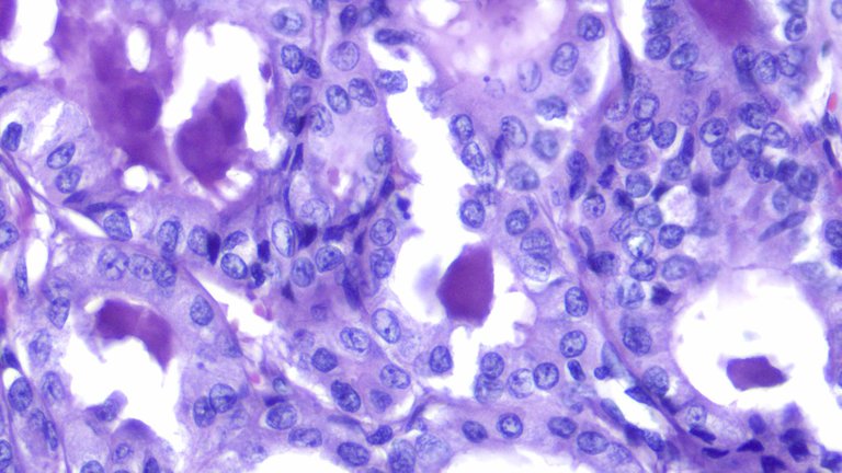
Malignant cells don’t respect personal space that’s why they overlap. Some look like round cells being folded longitudinally (nuclear grooving), and the chromatin inside the nuclei can be optically clear (Orphan-Annie). The staging and prognosis is favorable for people less than 55 years of age and prompt treatment.
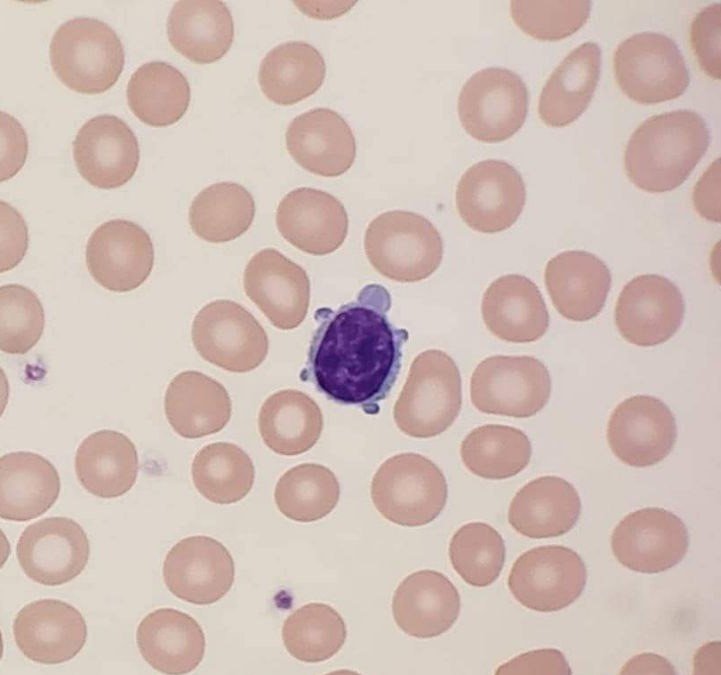
Look at this turtle looking lymphocyte on a peripheral blood smear, looked funny.
If you made it this far reading, thank you for your time.
Posted with STEMGeeks
Your content has been voted as a part of Encouragement program. Keep up the good work!
Use Ecency daily to boost your growth on platform!
Support Ecency
Vote for new Proposal
Delegate HP and earn more
We can see clearly the difference, do you think that this is one of the easiest ones to differentiate from a normal tissue behavior for a not trained eye?

!1UP
The differences here are on easy mode given that it's from tissue sections. When we get aspirate specimens, these can be difficult to appreciate especially for bloody smears. The classic features may not be readily distinguishable so further resampling may be recommended.
Tissue biopsy is recommended but the choice to operate isn't something to be taken lightly as one could require only one lobe to remove or two depending on the extent of tumor involvement on imaging.
You have received a 1UP from @gwajnberg!
@stem-curator
And they will bring !PIZZA 🍕.
Learn more about our delegation service to earn daily rewards. Join the Cartel on Discord.
Thanks for your contribution to the STEMsocial community. Feel free to join us on discord to get to know the rest of us!
Please consider delegating to the @stemsocial account (85% of the curation rewards are returned).
You may also include @stemsocial as a beneficiary of the rewards of this post to get a stronger support.
Haha, you found the turtle.
!discovery 37
This post was shared and voted inside the discord by the curators team of discovery-it
Join our community! hive-193212
Discovery-it is also a Witness, vote for us here
Delegate to us for passive income. Check our 80% fee-back Program
Congratulations @adamada.stem! You have completed the following achievement on the Hive blockchain and have been rewarded with new badge(s):
Your next target is to reach 15000 upvotes.
You can view your badges on your board and compare yourself to others in the Ranking
If you no longer want to receive notifications, reply to this comment with the word
STOPCheck out the last post from @hivebuzz:
Support the HiveBuzz project. Vote for our proposal!