Mucinous Cystadenocarcinoma Show and Tell
Sharing you one of the cases I handled in the past.
This was a case of a 47 year old, female that was managed as a case of ovarian carcinoma stage 4B. I don’t want to turn this post into a boring lecture as I just want to highlight the images from histology. We recently got our 5-header microscope fixed but the cables for the digital camera need some replacement, this makes my show and tell via pictures limited in quality.
The findings I have for this case focusing only on the ovary sections:
Histologic findings show ovarian tissue invaded by malignant neoplasm arranged in well-formed to poorly formed glands, and occasionally shows comedo necrosis. and occasional stromal invasion. The tumor cells have vesicular, pleomorphic nuclei with prominent nucleoli and moderate to ample cytoplasm. Brisk mitotic activity is observed.
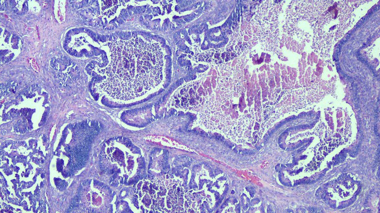
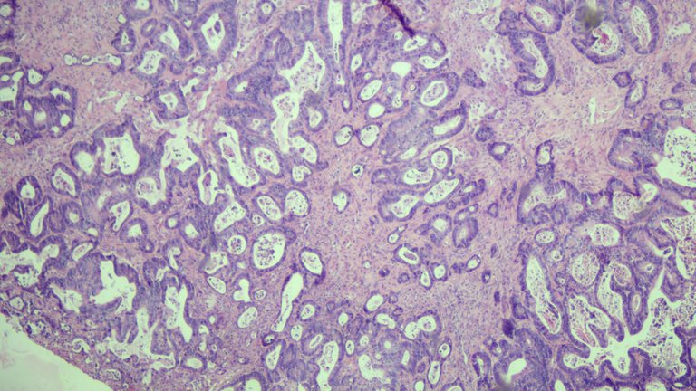
This was a section of the ovary taken at scanner view. You can see the entire architecture of the tumor forming well formed to poorly formed glands.
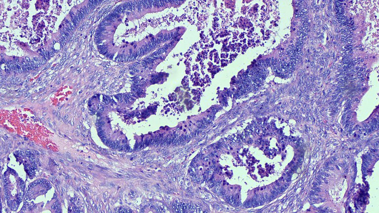
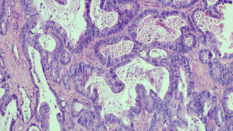
This was taken at 100x magnification. This view lets me survey the slide at closer details to spot where I should focus on. The center on the lumen shows necrotic debris (dead cells, tumor cells, inflammatory cells and etc.)
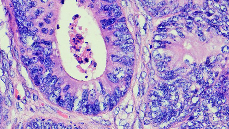
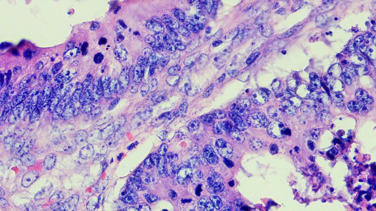
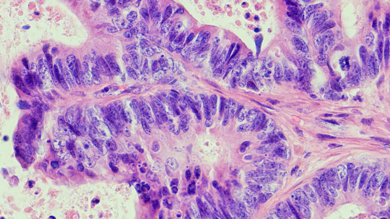
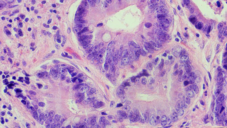
These two shots were taken at high power (400x magnification). This view is meant to distinguish fine cellular details like mitotic activity, chromatin pattern, and cytoplasmic inclusions just to name a few. Those bar like or round dense blue thingies can be cells undergoing mitosis.
I also include what the tumor looks like when it invaded other organs like the liver and rectum below. I didn’t bother putting in some pointers as the focus of the pictures are already bizarre tumor cells.
Taken at scanner view 40x magnification.
Taken at low power 100x magnification.
Taken at high power 400x magnification. The tumor form doesn't stray away from the morphology demonstrated from the tumor found in the ovary.
And some views from the sampled rectal tissues:
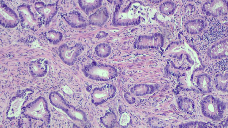
Taken at 100x magnification. The right side still has semblance to what a normal colonic mucosa would look like but the stroma which covers 3/4 left side of the image shows invasion.
Taken at high power. The morphology isn't that far from the main source of the tumor features.
I really like taking pictures of these tumors with the 5 header for more detail. I was tempted to make the post a lecture type but then I would just bore an audience. This concludes my show and tell.
If you made it this far reading, thank you for your time.
Posted with STEMGeeks
Congratulations @adamada.stem! You have completed the following achievement on the Hive blockchain and have been rewarded with new badge(s) :
Your next target is to reach 4000 upvotes.
You can view your badges on your board and compare yourself to others in the Ranking
If you no longer want to receive notifications, reply to this comment with the word
STOPTo support your work, I also upvoted your post!
Check out the last post from @hivebuzz:
Support the HiveBuzz project. Vote for our proposal!
I see we work in the same field....
Pathology specifically or another branch of medicine?