Krukenberg Tumors
Krukenberg Tumors are malignancies that involve the ovaries from cancers coming from the gastrointestinal system, the most common organ source would be the stomach. Here's a gross look at the ovaries taken out from the patient on their late 40s.
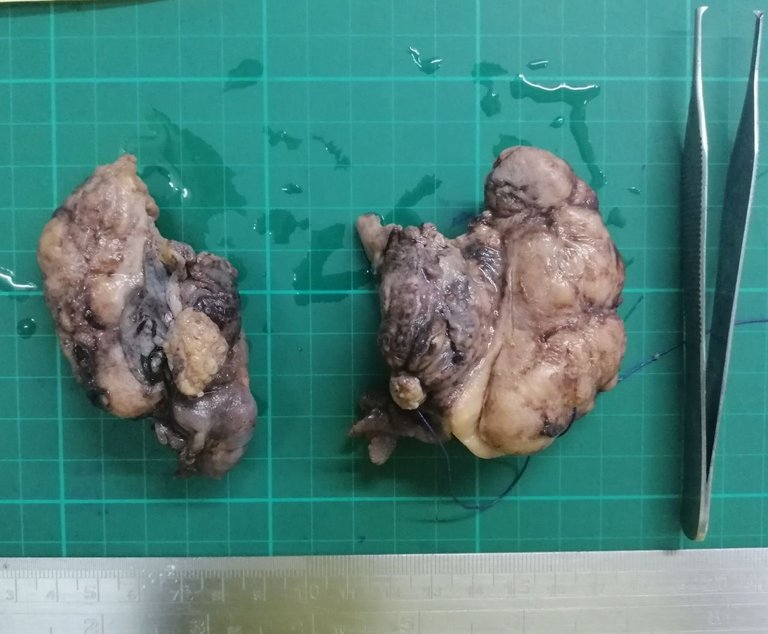
Signs of the tumor being metastatic grossly:
- The ovarian surface looks like it has a lot of implants
- Bilateral involvement
- Multiple organs suspected involved.
On this case, the manifestations were confined to the ovaries. These tumors often spell poor prognosis because their manifestations are discovered when they have already spread, on this case, its the patient's ovaries. I wasn't able to take photo the cut surface but Krukenberg tumors are fairly common enough. Like most stuff I post, anyone can just Google the medical related content. My posts are just written for show and tell.
Taken at scanner, low power view, and high power view respectively:


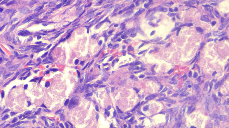
The large granular pink (eosinophilic) to clear cells look like signet ring with the hyperchromatic nucleus displaced at the periphery infiltrating the ovarian tissue arranged in sheets.
Metastatic tumor cells don't necessarily appear like their source tumors but when they accumulate in another site, they may form structures similar to the primary tumor.
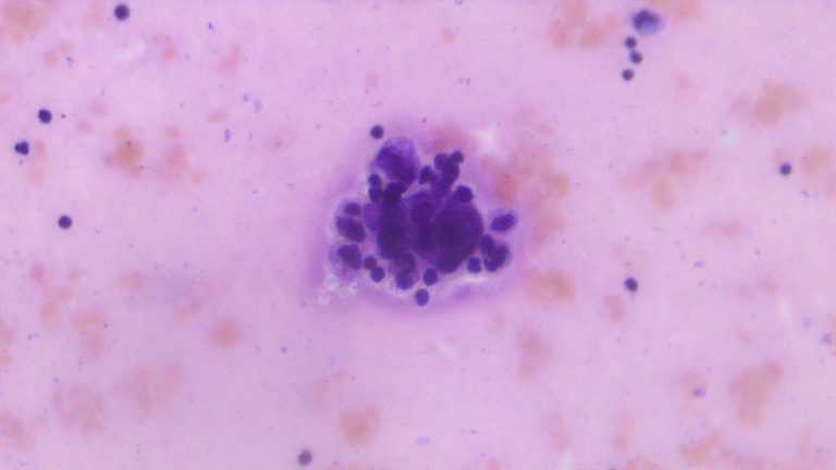
Here's a cluster of tumor cells from the patient's peritoneal fluid:
Tumors cells invading the omentum in the middle taken at low power view:
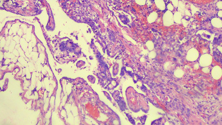
Tumor involving the peritoneum at low power view and high power view:
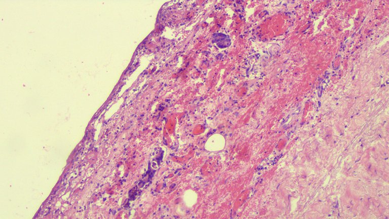
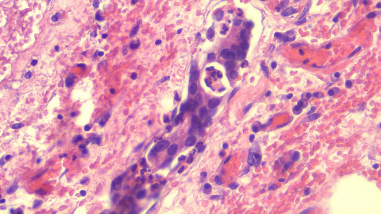
I wasn't able to take a photo of the immunohistochemical studies done on this patient but it turned out positive for CK7,CK20, CA19-9 and negative for Mammoglobin. This means other than confirming the metastatic cells came from the Gastrointestinal Organs, it's also hinting that stomach or pancreas may be the primary source. The negative mammoglobin was just trying to rule out the malignancy came from the breast.
The patient died before they could perform an endoscopic exam to check the duodenal area. No autopsy was requested to confirm the source. This has been a prevailing problem with the culture here as folks tend to see autopsies as one way of not letting the dead be laid to rest and prolonging the agony.
It's the first time I encountered this case and it wasn't a hard diagnosis to come up with. The next differential diagnosis includes the benign Signet Ring Stromal Tumors and related Sertoli-Leydig Tumors. Seeing the other specimens positive for tumor was more than enough to clinch the diagnosis.
If you made it this far reading, thank you for your time.
Posted with STEMGeeks
RIP.
Do you see yourself getting better at grossing now?
!discovery 41
Doubt, it's been 4-5 months since the last time I cut anything. These were just old cases I did and retrieved for documentation. We have case conferences where they bring up old cases from the past and I just take photographs for powerpoints, side perk is getting something to post about. I should be worried considering my evaluation from consultants said I suck at grossing specimens. Looking forward to December where I'm back at cutting stuff again.
Oof. Good luck with that.
This post was shared and voted inside the discord by the curators team of discovery-it
Join our community! hive-193212
Discovery-it is also a Witness, vote for us here
Delegate to us for passive income. Check our 80% fee-back Program
So sad! Maybe she took to much time to seek help?
!1UP
That and partly because these tumors tend to be insidious only showing up when it's already spread out.
You have received a 1UP from @gwajnberg!
@stem-curator
And they will bring !PIZZA 🍕.
Learn more about our delegation service to earn daily rewards. Join the Cartel on Discord.
Thanks for your contribution to the STEMsocial community. Feel free to join us on discord to get to know the rest of us!
Please consider delegating to the @stemsocial account (85% of the curation rewards are returned).
You may also include @stemsocial as a beneficiary of the rewards of this post to get a stronger support.
Ew those don't look fantastic.
These images make me kind of want to get a microscope again as my little walking hazard is now plenty old enough to not destroy one (inadvertently or otherwise) but I have a storage issue -_-
Storage for the microscope or the slides used? slides only last around 3 years before you notice a drop in quality unless they were already processed badly. Using the microscope for photography is fun, the downside is camera's attached are expensive and it's a niche content zone.
The microscope. The slides would (probably) survive.
While Krukenberg tumors can occur in women of any age, they are most often diagnosed in postmenopausal women. These tumors tend to be larger at the time of diagnosis and are often more advanced than other types of ovarian cancer. Krukenberg tumors often have a poor prognosis, but treatment options are available and the outlook can vary depending on the individual case.
Thank you for this great info
Not sure if the comment was for me or just someone looking at the post? if for me, yeah I know, it's my case.
I wonder why it's very hard for people living with Krukenberg Tumors as research has it that only 10% of patients survive after two years of diagnosis.
Because these manifestations of another primary cancer which has already spread and hard to treat during the time of diagnosis.
Congratulations @adamada.stem! You have completed the following achievement on the Hive blockchain And have been rewarded with New badge(s)
Your next target is to reach 9000 upvotes.
You can view your badges on your board and compare yourself to others in the Ranking
If you no longer want to receive notifications, reply to this comment with the word
STOPCheck out the last post from @hivebuzz:
Support the HiveBuzz project. Vote for our proposal!
Those ovaries looks like something off the side of a boat.