Found some T. Vaginalis on Pap Smears
Sharing some learning experience with pap smears. These tests take a swab sample from the cervix to screen for cervical lesions that may manifest the early signs of malignancy. There's plenty of stuff shared on Google that can explain the procedure well and that's not the point of this post.
TL:DR the pictures on the book are nice but nothing beats experience. Despite how much I'm not attached to my work, I'm still glad that I get to encounter novel or difficult cases as much as I can. It's difficult to ask for help once you're a step above the ladder where people expect you already mastered something you didn't have the chance to learn from while training.
Just sharing a learning experience related to textbook ideas versus what it actually is in practice. I had the mental image that Trichomas vaginalis would be easy to identify under a still image much like a textbook or Google images make it distinct to be but when my consultant pointed out I missed the pathology when reviewing the slide this was when the lesson started.
Taken at high power view (400x)
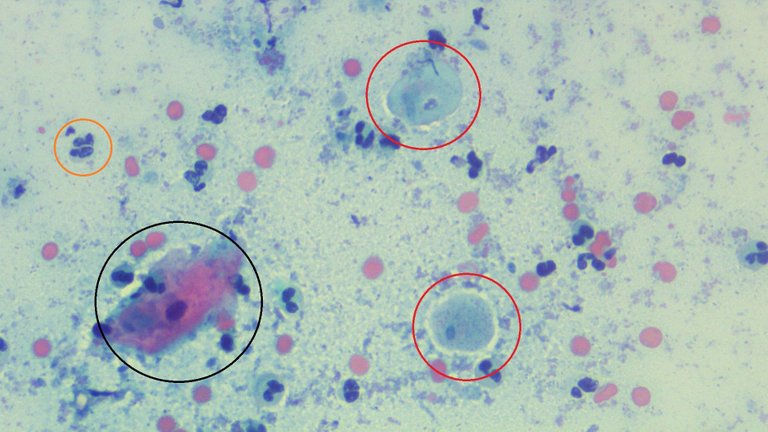
Yellow circle: A neutrophil
Black circle: An epithelial cell, intermediate.
Red Circle: T. vaginalis which I initially thought it was just a poor stained intermediate cell given the color.
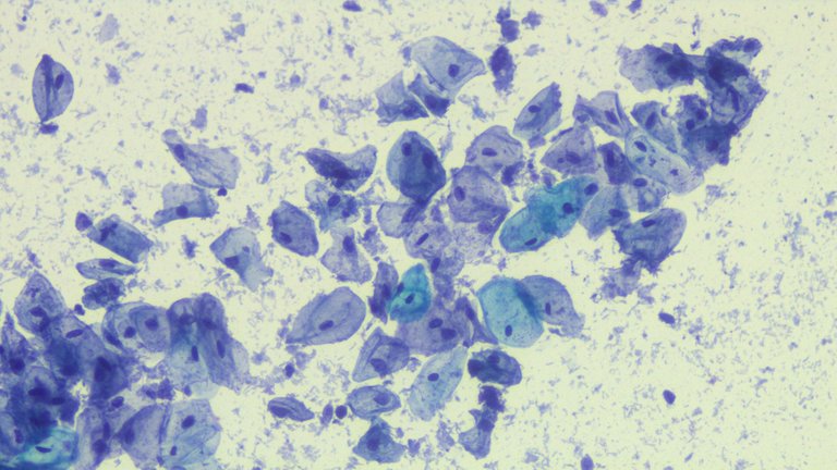
Taken at Low Power View (100x), these are intermediate cells surrounded by debris.
If you haven't noticed the subtle contrast, here's what you missed:
- T. vaginalis looks like it has a halo
- The nuclei is paler compared to the nuclei of intermediate cells.
- The nuclei is grooved on intermediate cells and the epithelial cells are slightly larger than T. vaginalis.
The pitfall from not recognizing this was getting used to textbook images that display perfect preservation of the features of the pathogen, like a distinct flagella.
I'm used to reading bad smears. It's not like I can control operator technique if the specimen was collected by inexperienced hands or bad preparation was done. The second image above is the norm but if you check out Google images for pap smear epithelial cells, you'll occasionally see "clean" fields.
Had this been prepared on a wet mount where the organism was still alive doing it's classic falling leaf motion, it would have been easy to spot. Despite these pathogens being reported common on paper, this is the first I've encountered a case and by luck, the second that followed was now easy to spot.
A consultant commented that once you recognize 1 T. vaginalis in the slide, you'll eventually start seeing a lot of them around. I have a habit of not overdoing the diagnosis just to keep the confirmation bias at a manageable level.
One piece of wisdom when it comes to diagnosing cases is that if you're fixated on looking for a pathology, you'll likely find one when there isn't. My rates of calling something malignant when benign dropped after reflecting on this piece of advice.
If you made it this far reading, thank you for your time.
Posted with STEMGeeks
Thanks for your contribution to the STEMsocial community. Feel free to join us on discord to get to know the rest of us!
Please consider delegating to the @stemsocial account (85% of the curation rewards are returned).
You may also include @stemsocial as a beneficiary of the rewards of this post to get a stronger support.
it is common in to find it! but good job. The problem is when you have " infestation"

!1UP
You have received a 1UP from @gwajnberg!
@stem-curator
And they will bring !PIZZA 🍕.
Learn more about our delegation service to earn daily rewards. Join the Cartel on Discord.