Clear Cell Neoplasms from Different Organ of Origins
Showcasing how clear cell carcinomas look across different organs involved. Please note that this is a phenotypic expression where neoplastic cells have a characteristic hyperchromatic central to eccentric nuclei and abundant clear cytoplasm. These can have a different aberration at a genetic and molecular level but the end expression makes them look like clear cells. I only have three organs for this post from Ovaries, Kidney and Brain specimens.
I'm not going to go into details about how these came to be or prognosis. The post is simply sharing how the same characteristic of neoplastic cells are visibly the almost the same and yet found on different organ locations bearing different clinical implications.
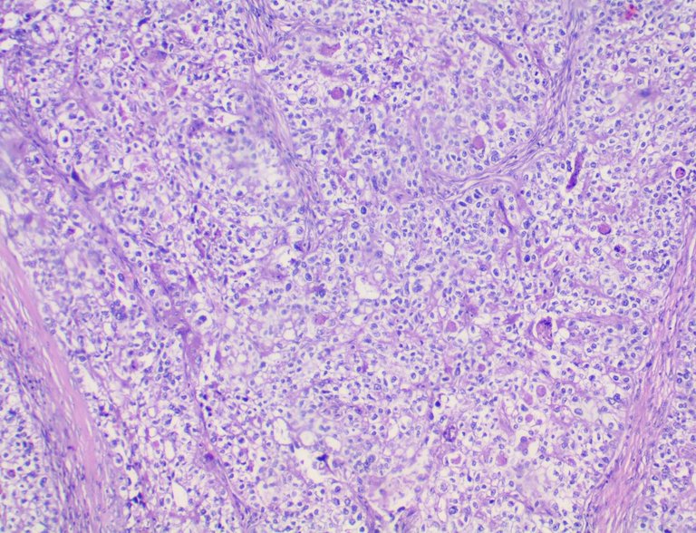
Clear Cell Carcinoma of the Ovary
Taken at Low Power View (100x):
Taken at High Power View (400x)
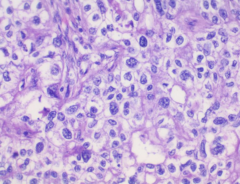
I've never encountered this case before and the pictures came from a colleague's case which I borrowed for study.
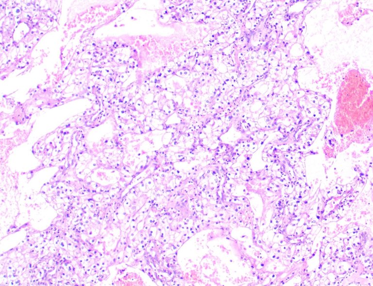
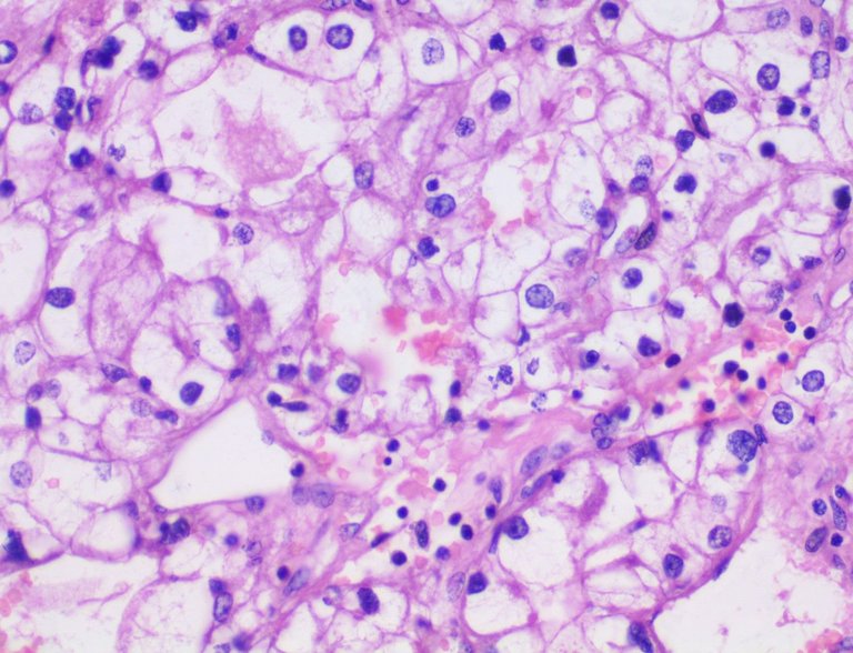
Renal Clear Cell Carcinoma
Taken at Low Power Views (100x)
Taken an High Power View (400x)
I don't get to cut a lot of radical nephrectomy specimens so I've only seen two cases with renal clear cell carcinoma twice in my training.
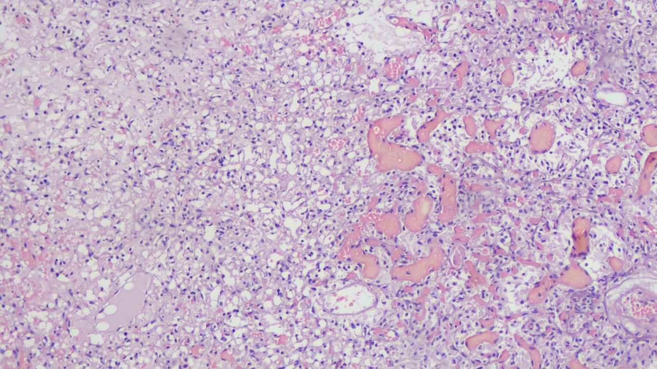
A throwback on the Hemangioblastoma case with clear cells.
Taken at Low Power View (100x)
Taken at High Power View (400x)
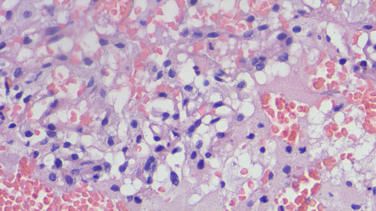
I mentioned before that committing to Hemangioblastoma requires ruling out possible metastatic renal clear cell carcinoma metastasis because you can see the Renal Clear Cell Carcinoma above as an example of how morphology isn't enough sometimes.
Now imagine receiving a slide looking at these features and then no one tells you the origin of the specimen?
If you made it this far reading, thank you for your time.
Posted with STEMGeeks
Congratulations @adamada.stem! You have completed the following achievement on the Hive blockchain and have been rewarded with new badge(s):
Your next target is to reach 10000 upvotes.
You can view your badges on your board and compare yourself to others in the Ranking
If you no longer want to receive notifications, reply to this comment with the word
STOPCheck out the last post from @hivebuzz:
Support the HiveBuzz project. Vote for our proposal!
Interesting how at 400x it becomes more clear what is happening!
!1UP
<a href="https://discord.gg/zQrvxAu7mu"> <img src="https://files.peakd.com/file/peakd-hive/thecuriousfool/23wCNFDyCLJu1v77TTg2MYKkd7XWkgF9fhiLujTDAaLaUz7H4AaQkDentB5UMVS8FcrVs.png"></a>You have received a 1UP from @gwajnberg!
@stem-curator
And they will bring !PIZZA 🍕.
Learn more about our delegation service to earn daily rewards. Join the Cartel on Discord.
I know that all too well. I get specimens labeled "swabs" for sources here and there. The floor seems to have trouble understanding the source affects the diagnosis greatly when we recover organisms.
!discovery 37
Ikr? sometimes we receive pieces of flesh or aspirate smears where it takes hours to days worth of delay just to get in contact with the folks/institution that sent it. This tiny detail really grinds my gears as I have to go through different people just to search for the one that sent the specimen in the lab for reference.
Literally affects patient care.
This post was shared and voted inside the discord by the curators team of discovery-it
Join our community! hive-193212
Discovery-it is also a Witness, vote for us here
Delegate to us for passive income. Check our 80% fee-back Program
Thanks for your contribution to the STEMsocial community. Feel free to join us on discord to get to know the rest of us!
Please consider delegating to the @stemsocial account (85% of the curation rewards are returned).
You may also include @stemsocial as a beneficiary of the rewards of this post to get a stronger support.