Artifacts and Tumor Cells
Cytology smears have always been a source of frustration during slide review. The problem with doing this is how much time one has to use just viewing each slide. It's directly proportional to experience where the more you get used to it the less time you spent looking at it.
Now between a consultant's eye and someone in training, the spells a huge gap in skill and time saved looking at the slides. Below is a slide processed from the peritoneal fluid of an ovarian malignancy I previously suspected here.
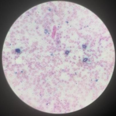
Part of the confirmation bias is trying to find something unusual because you believe there is. Now I looked at these and find it odd to see something resembling an calcium oxalate crystals on peritoneal fluid. I mean, maybe it's possible because having calcified degeneration also mean hardened calcium deposits but nope, this is just an artifact from a broken cover slip. It's a rookie mistake.
This is from a different patient and it's pleural fluid.
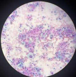
You can tell these are not normal cells based on the lack of uniformity on their nuclear sizes. Some white blood cells and other debris are in the mix but the large cells give it away making this sample positive for tumor cells. It was an easy slide.
After being bombarded with series of exams, take home work, and all other requirements imposed by the system, I have come to a burned out state where I just recently submitted documents online containing my answers for an online exam with half unanswered and unmonitored.
The proctor knew I could just cheat the system easy and there was no way someone else could prove it of course. I just said fuck it and passed the papers after seeing Mycology wasn't my best subject. Too tired to care anymore about performance ratings and feedback, if they want to fire me for underperforming the academic parts of the training, I won't mind anymore.
If you made it this far reading, thank you for your time.
Posted with STEMGeeks
Haha, the hardest thing on any practical back when I was in school was always the normal specimens. It's very easy to trick students into thinking there's something wrong with the blood smear, chemistry panel, etc.
Oh and mycology. Very few people are good at it. I wouldn't lose sleep over it. It's a subject that you are either all in or just good enough to get by.
It's more or less like that while at work but tends to lean on yeah there's probably something wrong here otherwise they won't send us this specimen in the first place bias.
Just got frustrated that I studied the agar formulas and their purpose only to be asked different questions often of the least priority. Like being told you study chapter 1 for the quiz as pointers but found out chapter 3 had all the answers. Got over it now~
When you have a minute, could you stick some arrows on the second slide pointing out what all the bits are please?
I think I got your debris on the first slide, the needle shaped pink crystal?
Keep em coming you two!
Will note those arrows on future posts :P
The 1st slide, those 3 box shaped transparent crystals forming a triangle looked like calcium oxalate.
the second slide is the exact center where you see a cloud of pink cells made up of different smaller round and darker shade of pink circles. Those darker circles are their nucleus and they vary in sizes. If you look at the 7 o'clock position you would see a large round circle which has a space out around, that space is the cytoplasm and it looks "foamy" because it's a macrophage eating up the debris or reacting to the surrounding inflammation.
so the combined cells are tumors I didn't know of this before