A Case of Pineoblastoma
This came from a 4 year old female who presented with headache and gradual blurring of vision. MRI showed a pineal mass.
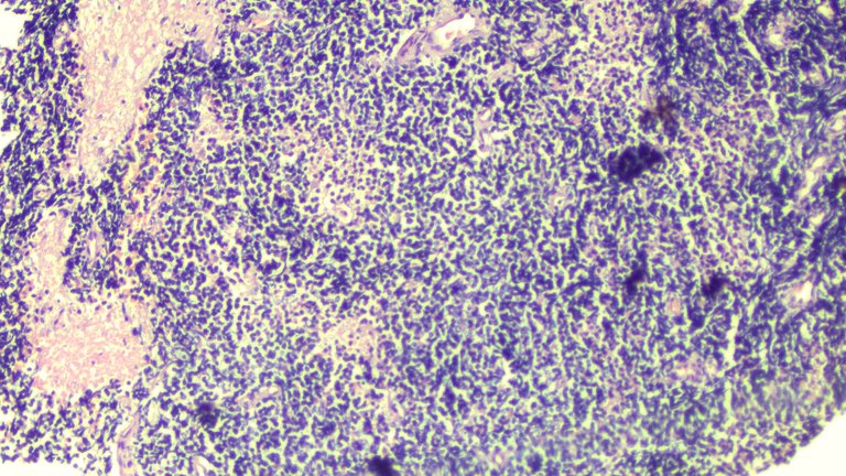
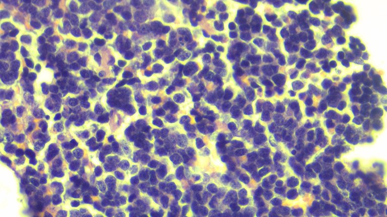
The tumor cells have a round to ovoid, hyperchromatic nuclei with inconspicuous nucleoli and scant cytoplasm. It's difficult to appreciate mitotic figures here but it's expected to be mitotically active. This is a Grade IV CNS tumor which immediately spells a worse prognosis. You can find a brief summary about it here
My images aren't the best so you're doing yourself a service googling up some examples of this case.
Low Power View:

High Power View:

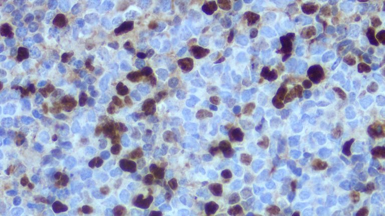
Ki-67 Stain at high power view:

The Ki-67 helps determine the proliferation index of the tumor, the higher the number the more active the tumor is multiplying. Since the common differentials for this case include Pineocytoma, Pineal Tumor of Intermediate Differentiation and Pineoblastoma, one could help rule out the others based on their mitotic index, with pineocytoma showing the least around less than 2%, to pineoblastoma above 20% then the pineal tumor of intermediate differentiation somewhere in between.
One term I throw around is "differentiation" as this implies how well the tumor resembles the normal tissue. The more well-differentiated it is, the better the prognosis but this is just a rule of thumb I make a mental note of and not absolute. Even if it's well-differentiated, a tumor can still spell a worse prognosis.
The morphology suggests a small round blue cell tumor, but this is the same energy as saying there's a lot of possible diagnosis we can come up with that fits the small round blue cell neoplasm spectrum so we need to use some immunohistochemical studies to help narrow down the lineage. The problem with tumors that present with small round blue cell morphology is that these can be seen as sarcomas, immature cells, or lymphomas. There are some clues to differentiate between them but in practice, it's not really worth sticking out your neck if there's no confirmatory test to back your claim.
I rarely encounter cases from the brain, especially if they present as classic textbook cases. It's fascinating but you know it's bad news for someone else. The patient expired shortly after this was removed. These tumors are aggressive and would have a poor prognosis from the start.
Other than Ki-67, we also ran SALL-4 to rule out germ cell tumors, CD45 to rule out lymphomas, NSE to rule in the diagnosis, GFAP to rule out other common differentials. I know I brushed over these stains and their specific purposes but stretching the post isn't exactly fun for the sake of trivia.
One thing to keep in mind with these cases is the diagnostic pitfall by just judging from morphology. Imaging studies are a must when it comes to clinching the diagnosis because another differential diagnosis for this case is medulloblastoma. If the location of the mass was far from the pineal region and near the cerebellar area, that would be the clue.
If you made it this far reading, thank you for your time.
Posted with STEMGeeks
Damn, died of cancer at 4 years old....
Some just don't have the luck with the genetic raffle.
Oh boy...this is sad and yet fascinating.
!discovery 43
It's a classic text book case. When I did a reporting on this just for academic didactics, I wasn't told about the full story so I just had to piece together the story from the stains used, and imaging without seeing the full report because patient privacy. When the consultant said they considered Medulloblastoma for this case that was horror as I spent the entire hour giving a lecture about Pineoblastoma only for them to say they signed out out as Pineoblastoma because imaging studies supported it.
Sounds like the evidence supported what was signed off.
This post was shared and voted inside the discord by the curators team of discovery-it
Join our Community and follow our Curation Trail
Discovery-it is also a Witness, vote for us here
Delegate to us for passive income. Check our 80% fee-back Program
Congratulations @adamada.stem! You have completed the following achievement on the Hive blockchain And have been rewarded with New badge(s)
Your next target is to reach 400 replies.
You can view your badges on your board and compare yourself to others in the Ranking
If you no longer want to receive notifications, reply to this comment with the word
STOPCheck out our last posts:
Thanks for your contribution to the STEMsocial community. Feel free to join us on discord to get to know the rest of us!
Please consider delegating to the @stemsocial account (85% of the curation rewards are returned).
You may also include @stemsocial as a beneficiary of the rewards of this post to get a stronger support.